· FCPS MCPS IMM MD Paediatrics TOACS
Observed Station | CPSP Format | 8 minutes
⏱️ TIME REMAINING
08:00
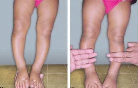
A 2‑year‑old child is brought to the clinic because of bowlegs (genu varum) that have been progressive over the past 6 months.
❓ Q1. Identify the radiographic finding shown in the image. What condition is it used to diagnose?
✅ Model Answer: • Radiographic finding: Cover-up test (Langenskiöld) – positive when the distal femoral condyle overlaps the tibial metaphysis on a lateral knee radiograph.
• Condition: Blount disease (tibia vara).
• The test helps differentiate Blount disease from physiologic genu varum (which has a negative cover-up test).
• Condition: Blount disease (tibia vara).
• The test helps differentiate Blount disease from physiologic genu varum (which has a negative cover-up test).
❓ Q2. What is Blount disease? What are its subtypes?
✅ Model Answer: • Blount disease (tibia vara): A growth disorder of the proximal tibial physis (medial physis) leading to progressive varus angulation, internal tibial rotation, and lateral thrust on gait.
• Subtypes:
- Infantile Blount disease: Onset before age 3 years. Often bilateral, associated with obesity and early walking.
- Juvenile Blount disease: Onset between 4-10 years.
- Adolescent Blount disease: Onset after age 10 years.
• Pathophysiology: Excessive mechanical load on the medial proximal tibial physis → growth suppression → varus deformity.
• Subtypes:
- Infantile Blount disease: Onset before age 3 years. Often bilateral, associated with obesity and early walking.
- Juvenile Blount disease: Onset between 4-10 years.
- Adolescent Blount disease: Onset after age 10 years.
• Pathophysiology: Excessive mechanical load on the medial proximal tibial physis → growth suppression → varus deformity.
❓ Q3. How is the cover-up test performed? What does a positive test indicate?
✅ Model Answer: • Technique: A lateral radiograph of the knee is obtained with the knee in full extension.
• The examiner observes the relationship between the distal femoral condyle and the proximal tibia.
• Positive test: The distal femoral condyle overlaps the tibial metaphysis (the femur "covers" the tibia). This indicates tibial torsion and is seen in Blount disease.
• Negative test: No overlap between the femur and tibia – seen in physiologic genu varum.
• The test is also called the Langenskiöld test.
• The examiner observes the relationship between the distal femoral condyle and the proximal tibia.
• Positive test: The distal femoral condyle overlaps the tibial metaphysis (the femur "covers" the tibia). This indicates tibial torsion and is seen in Blount disease.
• Negative test: No overlap between the femur and tibia – seen in physiologic genu varum.
• The test is also called the Langenskiöld test.
❓ Q4. What are the clinical features of infantile Blount disease?
✅ Model Answer: • Clinical features:
- Age: Usually presents between 1-3 years of age.
- Genu varum (bowlegs): Progressive, often bilateral.
- Obesity: Common risk factor (BMI >95th percentile).
- Early walking: Often begins walking early.
- Lateral thrust: A visible thrust of the knee laterally during gait (due to knee instability).
- Internal tibial torsion: The tibia rotates inward.
- Pain: Usually absent in early stages.
- Examination: Varus deformity, prominent proximal tibia (metaphyseal beak), lateral thrust on walking.
- Age: Usually presents between 1-3 years of age.
- Genu varum (bowlegs): Progressive, often bilateral.
- Obesity: Common risk factor (BMI >95th percentile).
- Early walking: Often begins walking early.
- Lateral thrust: A visible thrust of the knee laterally during gait (due to knee instability).
- Internal tibial torsion: The tibia rotates inward.
- Pain: Usually absent in early stages.
- Examination: Varus deformity, prominent proximal tibia (metaphyseal beak), lateral thrust on walking.
❓ Q5. What is the metaphyseal-diaphyseal angle (MDA)? How is it used in the diagnosis of Blount disease?
✅ Model Answer: • Metaphyseal-diaphyseal angle (MDA) – Levine and Drennan: The angle between a line drawn along the metaphysis of the proximal tibia and a line along the diaphysis of the tibia.
• Measured on an anteroposterior (AP) radiograph of the knee.
• Interpretation:
- <9°: Physiologic genu varum (normal).
- 9-11°: Borderline – observe closely.
- >11°: Diagnostic of Blount disease (pathologic).
• The MDA is a key radiographic parameter used to differentiate Blount disease from physiologic bowing.
• Measured on an anteroposterior (AP) radiograph of the knee.
• Interpretation:
- <9°: Physiologic genu varum (normal).
- 9-11°: Borderline – observe closely.
- >11°: Diagnostic of Blount disease (pathologic).
• The MDA is a key radiographic parameter used to differentiate Blount disease from physiologic bowing.
❓ Q6. What is the Langenskiöld classification of Blount disease?
✅ Model Answer: • Langenskiöld classification (6 stages): A radiographic staging system for infantile Blount disease based on progressive metaphyseal changes.
- Stage I (0-2 years): Irregularity and ossification defects of the medial metaphysis.
- Stage II (2-3 years): Metaphyseal beaking.
- Stage III (3-4 years): Progressive beaking and fragmentation.
- Stage IV (4-5 years): Severe beaking with physeal involvement.
- Stage V (6-8 years): Medial plateau depression (double contour).
- Stage VI (8-10 years): Bony bridging of the physis (complete growth arrest).
• Clinical utility: Guides treatment decisions – early stages (I-II) may be managed with bracing; advanced stages (III-VI) require surgery.
- Stage I (0-2 years): Irregularity and ossification defects of the medial metaphysis.
- Stage II (2-3 years): Metaphyseal beaking.
- Stage III (3-4 years): Progressive beaking and fragmentation.
- Stage IV (4-5 years): Severe beaking with physeal involvement.
- Stage V (6-8 years): Medial plateau depression (double contour).
- Stage VI (8-10 years): Bony bridging of the physis (complete growth arrest).
• Clinical utility: Guides treatment decisions – early stages (I-II) may be managed with bracing; advanced stages (III-VI) require surgery.
❓ Q7. How does Blount disease differ from physiologic genu varum?
✅ Model Answer: • Blount disease:
- Age: >2 years (infantile type).
- Progression: Progressive, worsens with growth.
- Obesity: Common.
- Lateral thrust: Present.
- Cover-up test: Positive (femoral overlap).
- MDA: >11°.
- Treatment: Bracing or surgery required.
• Physiologic genu varum:
- Age: 0-2 years (resolves by age 2-3 years).
- Progression: Self-limiting, resolves with growth.
- Obesity: Not typical.
- Lateral thrust: Absent.
- Cover-up test: Negative (no overlap).
- MDA: <9°.
- Treatment: Observation only.
- Age: >2 years (infantile type).
- Progression: Progressive, worsens with growth.
- Obesity: Common.
- Lateral thrust: Present.
- Cover-up test: Positive (femoral overlap).
- MDA: >11°.
- Treatment: Bracing or surgery required.
• Physiologic genu varum:
- Age: 0-2 years (resolves by age 2-3 years).
- Progression: Self-limiting, resolves with growth.
- Obesity: Not typical.
- Lateral thrust: Absent.
- Cover-up test: Negative (no overlap).
- MDA: <9°.
- Treatment: Observation only.
❓ Q8. What is the management of infantile Blount disease?
✅ Model Answer: • Management based on stage and age:
- Mild disease (Langenskiöld I-II, age <3 years): Knee-ankle-foot orthosis (KAFO) with a medial upright. Worn 23 hours/day for 1-2 years. Goal: correct the varus and allow growth.
- Moderate to severe disease (Langenskiöld III+, age >3 years):
- Guided growth (hemiepiphysiodesis): Tension band plate (eight-plate) on the lateral side of the proximal tibia to stimulate growth on the lateral side and correct the varus.
- High tibial osteotomy (HTO): For severe or advanced disease (Langenskiöld V-VI) or when growth arrest has occurred.
- Obesity management: Weight loss is essential to reduce mechanical load and improve outcomes.
- Mild disease (Langenskiöld I-II, age <3 years): Knee-ankle-foot orthosis (KAFO) with a medial upright. Worn 23 hours/day for 1-2 years. Goal: correct the varus and allow growth.
- Moderate to severe disease (Langenskiöld III+, age >3 years):
- Guided growth (hemiepiphysiodesis): Tension band plate (eight-plate) on the lateral side of the proximal tibia to stimulate growth on the lateral side and correct the varus.
- High tibial osteotomy (HTO): For severe or advanced disease (Langenskiöld V-VI) or when growth arrest has occurred.
- Obesity management: Weight loss is essential to reduce mechanical load and improve outcomes.
❓ Q9. What is the role of bracing in Blount disease?
✅ Model Answer: • Role of bracing:
- Indications: Infantile Blount disease, Langenskiöld stage I or II, in children <3 years old.
- Type: Knee-ankle-foot orthosis (KAFO) with a medial upright and a valgus-producing strap.
- Wear schedule: 23 hours/day for 12-18 months.
- Goal: Correct the varus deformity by applying a valgus force on the proximal tibia while allowing growth.
- Success: Bracing is successful in >70% of stage I cases, but less effective in stage II or older children.
- Monitoring: Serial radiographs (MDA, Langenskiöld stage) to assess response.
- Indications: Infantile Blount disease, Langenskiöld stage I or II, in children <3 years old.
- Type: Knee-ankle-foot orthosis (KAFO) with a medial upright and a valgus-producing strap.
- Wear schedule: 23 hours/day for 12-18 months.
- Goal: Correct the varus deformity by applying a valgus force on the proximal tibia while allowing growth.
- Success: Bracing is successful in >70% of stage I cases, but less effective in stage II or older children.
- Monitoring: Serial radiographs (MDA, Langenskiöld stage) to assess response.
❓ Q10. What is guided growth (hemiepiphysiodesis) in Blount disease?
✅ Model Answer: • Guided growth (hemiepiphysiodesis): A minimally invasive surgical procedure to correct angular deformities in growing children.
• Mechanism: A tension band plate (eight-plate) is placed on the lateral side of the proximal tibial physis. This slows growth on the lateral side while the medial side continues to grow, gradually correcting the varus deformity.
• Indications: Langenskiöld stage III or higher, or age >3 years with progressive deformity.
• Advantages: Less invasive than osteotomy, allows gradual correction, preserves growth potential.
• Monitoring: Regular radiographs to assess correction; plate removed when neutral alignment is achieved.
• Risks: Hardware failure, overcorrection, physeal arrest.
• Mechanism: A tension band plate (eight-plate) is placed on the lateral side of the proximal tibial physis. This slows growth on the lateral side while the medial side continues to grow, gradually correcting the varus deformity.
• Indications: Langenskiöld stage III or higher, or age >3 years with progressive deformity.
• Advantages: Less invasive than osteotomy, allows gradual correction, preserves growth potential.
• Monitoring: Regular radiographs to assess correction; plate removed when neutral alignment is achieved.
• Risks: Hardware failure, overcorrection, physeal arrest.
❓ Q11. What is the role of high tibial osteotomy (HTO) in Blount disease?
✅ Model Answer: • High tibial osteotomy (HTO): A surgical procedure to correct severe varus deformity by cutting and repositioning the proximal tibia.
• Indications:
- Langenskiöld stage V or VI (severe, with growth arrest).
- Children >8 years of age.
- Failure of bracing or guided growth.
- Severe varus with medial plateau depression.
• Technique: Osteotomy of the proximal tibia (valgus-producing), fixation with a plate or external fixator.
• Advantages: Provides immediate correction, addresses medial plateau depression.
• Risks: Compartment syndrome, neurovascular injury, nonunion, overcorrection.
• Indications:
- Langenskiöld stage V or VI (severe, with growth arrest).
- Children >8 years of age.
- Failure of bracing or guided growth.
- Severe varus with medial plateau depression.
• Technique: Osteotomy of the proximal tibia (valgus-producing), fixation with a plate or external fixator.
• Advantages: Provides immediate correction, addresses medial plateau depression.
• Risks: Compartment syndrome, neurovascular injury, nonunion, overcorrection.
❓ Q12. What is the differential diagnosis of genu varum in children?
✅ Model Answer: • Differential diagnoses:
- Physiologic genu varum: Normal in children <2 years, resolves spontaneously.
- Blount disease (tibia vara): Progressive varus, obesity, lateral thrust, positive cover-up test, MDA >11°.
- Rickets: Vitamin D deficiency, calcium/phosphate abnormalities, widened physis (cupping and fraying on X-ray), bowing of long bones.
- Metabolic bone disease: Hypophosphatasia, renal osteodystrophy.
- Trauma: Physeal injury (growth arrest).
- Infection: Septic arthritis, osteomyelitis affecting the physis.
- Skeletal dysplasias: Achondroplasia, metaphyseal dysplasia.
- Rheumatologic: Juvenile idiopathic arthritis (JIA) – can cause growth disturbances.
- Physiologic genu varum: Normal in children <2 years, resolves spontaneously.
- Blount disease (tibia vara): Progressive varus, obesity, lateral thrust, positive cover-up test, MDA >11°.
- Rickets: Vitamin D deficiency, calcium/phosphate abnormalities, widened physis (cupping and fraying on X-ray), bowing of long bones.
- Metabolic bone disease: Hypophosphatasia, renal osteodystrophy.
- Trauma: Physeal injury (growth arrest).
- Infection: Septic arthritis, osteomyelitis affecting the physis.
- Skeletal dysplasias: Achondroplasia, metaphyseal dysplasia.
- Rheumatologic: Juvenile idiopathic arthritis (JIA) – can cause growth disturbances.
❓ Q13. How would you counsel the parents of a child with Blount disease?
✅ Model Answer: • "Your child has been diagnosed with Blount disease, a condition that affects the growth plate of the shin bone (tibia), causing the leg to bow outward. It is more common in children who are overweight and walk early."
• "We have confirmed the diagnosis with X-rays, including a special test called the cover-up test, which shows the femur overlapping the tibia."
• "Treatment depends on the severity and your child's age. For mild cases, we may recommend a special brace (KAFO) to correct the bowing. For more advanced cases, surgery such as guided growth or an osteotomy may be needed to straighten the leg."
• "Weight management is very important to reduce the stress on the knee and improve outcomes."
• "We will monitor your child regularly with X-rays and exams to ensure the treatment is working and to prevent long-term problems like arthritis."
• "We have confirmed the diagnosis with X-rays, including a special test called the cover-up test, which shows the femur overlapping the tibia."
• "Treatment depends on the severity and your child's age. For mild cases, we may recommend a special brace (KAFO) to correct the bowing. For more advanced cases, surgery such as guided growth or an osteotomy may be needed to straighten the leg."
• "Weight management is very important to reduce the stress on the knee and improve outcomes."
• "We will monitor your child regularly with X-rays and exams to ensure the treatment is working and to prevent long-term problems like arthritis."
❓ Q14. What is the prognosis for a child with Blount disease?
✅ Model Answer: • Prognosis: Depends on the age of onset, severity, and response to treatment.
- Early diagnosis and treatment (infantile, stage I): Bracing is successful in >70% of cases, with good correction and normal function.
- Advanced disease (stage III+): Surgery is often required. Guided growth has excellent outcomes with gradual correction. HTO provides good correction but has higher morbidity.
- Untreated or progressive Blount disease: Leads to severe varus, gait abnormalities, and early osteoarthritis (medial compartment) in adulthood.
- Obesity: Poorly controlled obesity is associated with worse outcomes and higher recurrence rates.
- Long-term follow-up: Even after treatment, patients need monitoring until skeletal maturity to ensure correction is maintained.
- Early diagnosis and treatment (infantile, stage I): Bracing is successful in >70% of cases, with good correction and normal function.
- Advanced disease (stage III+): Surgery is often required. Guided growth has excellent outcomes with gradual correction. HTO provides good correction but has higher morbidity.
- Untreated or progressive Blount disease: Leads to severe varus, gait abnormalities, and early osteoarthritis (medial compartment) in adulthood.
- Obesity: Poorly controlled obesity is associated with worse outcomes and higher recurrence rates.
- Long-term follow-up: Even after treatment, patients need monitoring until skeletal maturity to ensure correction is maintained.
❓ Q15. What is the role of obesity management in Blount disease?
✅ Model Answer: • Obesity management: Obesity is a major risk factor for Blount disease and is associated with more severe deformity and poor treatment outcomes.
- Mechanism: Increased mechanical load on the medial proximal tibial physis → growth suppression → varus progression.
- Interventions:
- Dietary modification: Caloric reduction, balanced nutrition.
- Physical activity: Encourage low-impact exercise (swimming, cycling) to improve muscle strength and reduce joint stress.
- Weight loss: Even moderate weight loss can reduce symptoms and improve response to bracing or surgery.
- Multidisciplinary approach: Involve pediatric nutritionists and endocrinologists.
- Impact: Weight loss before surgery reduces the risk of complications and improves surgical outcomes.
- Mechanism: Increased mechanical load on the medial proximal tibial physis → growth suppression → varus progression.
- Interventions:
- Dietary modification: Caloric reduction, balanced nutrition.
- Physical activity: Encourage low-impact exercise (swimming, cycling) to improve muscle strength and reduce joint stress.
- Weight loss: Even moderate weight loss can reduce symptoms and improve response to bracing or surgery.
- Multidisciplinary approach: Involve pediatric nutritionists and endocrinologists.
- Impact: Weight loss before surgery reduces the risk of complications and improves surgical outcomes.
📋 Mock Test Feedback
💡 Examiner's note: Compare your answers with model answers. In real TOACS, you would discuss these with the examiner.