FCPS MCPS IMM MD Paediatrics TOACS
Observed Station | CPSP Format | 8 minutes
⏱️ TIME REMAINING
08:00
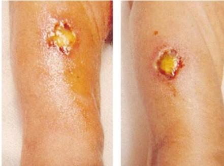
A 5‑day‑old term newborn in the NICU presents with fever, hypotension, and poor feeding. On examination, there are hemorrhagic bullae on the site of vit.K injection.
❓ Q1. Identify the skin condition shown in the image. What is the characteristic appearance of the lesions?
✅ Model Answer: • Condition: Ecthyma gangrenosum.
• Appearance: Starts as hemorrhagic bullae (fried egg appearance with necrotic center surrounded by inflammation) → progresses to necrotic ulcers with a black eschar. Lesions are often painless, rapidly progressive, and occur in neutropenic or immunocompromised patients. Common sites: perineum, axillae, trunk, extremities.
• Appearance: Starts as hemorrhagic bullae (fried egg appearance with necrotic center surrounded by inflammation) → progresses to necrotic ulcers with a black eschar. Lesions are often painless, rapidly progressive, and occur in neutropenic or immunocompromised patients. Common sites: perineum, axillae, trunk, extremities.
❓ Q2. Which organism is most commonly associated with ecthyma gangrenosum? What is the pathogenesis?
✅ Model Answer: • Organism: Pseudomonas aeruginosa (most common).
• Pathogenesis: Hematogenous spread of Pseudomonas → bacterial proliferation in the dermis → vasculitis with thrombosis and necrosis → hemorrhagic bullae → ulceration with black eschar.
• Occurs almost exclusively in neutropenic, immunocompromised, or septic patients.
• Other organisms: Aeromonas, Serratia, Klebsiella, E. coli (less common).
• Pathogenesis: Hematogenous spread of Pseudomonas → bacterial proliferation in the dermis → vasculitis with thrombosis and necrosis → hemorrhagic bullae → ulceration with black eschar.
• Occurs almost exclusively in neutropenic, immunocompromised, or septic patients.
• Other organisms: Aeromonas, Serratia, Klebsiella, E. coli (less common).
❓ Q3. What are the risk factors for developing ecthyma gangrenosum in neonates?
✅ Model Answer: • Risk factors:
- Neutropenia (absolute neutrophil count <500).
- Central venous catheter – common source of Pseudomonas.
- Prolonged antibiotic therapy (broad-spectrum antibiotics selecting for Pseudomonas).
- Prolonged NICU stay.
- Immunocompromise (neonates have immature immune systems).
- Recent surgery or invasive procedures.
- Congenital immunodeficiency (e.g., severe combined immunodeficiency).
- Neutropenia (absolute neutrophil count <500).
- Central venous catheter – common source of Pseudomonas.
- Prolonged antibiotic therapy (broad-spectrum antibiotics selecting for Pseudomonas).
- Prolonged NICU stay.
- Immunocompromise (neonates have immature immune systems).
- Recent surgery or invasive procedures.
- Congenital immunodeficiency (e.g., severe combined immunodeficiency).
❓ Q4. What is the classic lesion progression of ecthyma gangrenosum?
✅ Model Answer: • Progression (classic):
1️⃣ Stage 1 (Hours): Erythematous macule or papule → hemorrhagic bulla (appears as a "fried egg" – necrotic center with surrounding erythema).
2️⃣ Stage 2 (12-24 hours): Bulla ruptures → central ulceration.
3️⃣ Stage 3 (24-48 hours): Ulcer becomes necrotic with a black eschar (gangrenous center) surrounded by an erythematous halo.
4️⃣ Stage 4 (Days): Lesion may heal with scarring after debridement and treatment.
• Key feature: Lesions are often painless (due to nerve necrosis) but may become painful if superinfected.
1️⃣ Stage 1 (Hours): Erythematous macule or papule → hemorrhagic bulla (appears as a "fried egg" – necrotic center with surrounding erythema).
2️⃣ Stage 2 (12-24 hours): Bulla ruptures → central ulceration.
3️⃣ Stage 3 (24-48 hours): Ulcer becomes necrotic with a black eschar (gangrenous center) surrounded by an erythematous halo.
4️⃣ Stage 4 (Days): Lesion may heal with scarring after debridement and treatment.
• Key feature: Lesions are often painless (due to nerve necrosis) but may become painful if superinfected.
❓ Q5. What is the relationship between ecthyma gangrenosum and septicemia?
✅ Model Answer: • Ecthyma gangrenosum is a skin manifestation of Pseudomonas septicemia.
• In >90% of cases, it is associated with bacteremia.
• Lesions result from hematogenous spread of Pseudomonas to the skin.
• Can also occur from direct inoculation (less common).
• Mortality: High (up to 50% in neonates) if untreated or delayed treatment.
• The presence of ecthyma gangrenosum is a marker of severe sepsis and indicates the need for immediate broad-spectrum antipseudomonal antibiotics.
• In >90% of cases, it is associated with bacteremia.
• Lesions result from hematogenous spread of Pseudomonas to the skin.
• Can also occur from direct inoculation (less common).
• Mortality: High (up to 50% in neonates) if untreated or delayed treatment.
• The presence of ecthyma gangrenosum is a marker of severe sepsis and indicates the need for immediate broad-spectrum antipseudomonal antibiotics.
❓ Q6. What is the diagnostic approach for a neonate with suspected ecthyma gangrenosum?
✅ Model Answer: • Diagnostic approach:
- Blood cultures: Essential – often positive for Pseudomonas.
- Skin lesion culture: Swab or biopsy of the ulcer/eschar for Gram stain and culture.
- Skin biopsy: If diagnosis is uncertain (shows vasculitis, thrombosis, and Gram-negative bacilli).
- Complete blood count: Assess for neutropenia.
- CRP, procalcitonin: Markers of sepsis.
- Echocardiogram: To rule out infective endocarditis (if indicated).
- Blood cultures: Essential – often positive for Pseudomonas.
- Skin lesion culture: Swab or biopsy of the ulcer/eschar for Gram stain and culture.
- Skin biopsy: If diagnosis is uncertain (shows vasculitis, thrombosis, and Gram-negative bacilli).
- Complete blood count: Assess for neutropenia.
- CRP, procalcitonin: Markers of sepsis.
- Echocardiogram: To rule out infective endocarditis (if indicated).
❓ Q7. What is the first-line antibiotic treatment for ecthyma gangrenosum in a neonate?
✅ Model Answer: • Empiric antibiotics: Broad-spectrum antipseudomonal coverage.
• Options:
- Cefepime (4th generation cephalosporin) – good Pseudomonas coverage.
- Meropenem (carbapenem) – excellent Pseudomonas coverage.
- Piperacillin-tazobactam – good Pseudomonas coverage.
- ± Aminoglycoside (gentamicin or amikacin) for synergy in severe cases.
- If MRSA risk: Add vancomycin.
• Duration: 10-14 days (or longer if associated osteomyelitis, endocarditis).
• Adjust according to culture and sensitivity.
• Options:
- Cefepime (4th generation cephalosporin) – good Pseudomonas coverage.
- Meropenem (carbapenem) – excellent Pseudomonas coverage.
- Piperacillin-tazobactam – good Pseudomonas coverage.
- ± Aminoglycoside (gentamicin or amikacin) for synergy in severe cases.
- If MRSA risk: Add vancomycin.
• Duration: 10-14 days (or longer if associated osteomyelitis, endocarditis).
• Adjust according to culture and sensitivity.
❓ Q8. What is the role of surgical management in ecthyma gangrenosum?
✅ Model Answer: • Surgical management:
- Debridement: Removal of necrotic tissue and eschar to prevent further spread and promote healing.
- Source control: Remove or exchange central venous catheter (if line-associated).
- Wound care: Dressing changes, wound irrigation, and possible skin grafting for large defects.
- Importance: Surgical debridement combined with antibiotics is essential for optimal outcomes.
- Timing: Early debridement within 24-48 hours of diagnosis.
- Debridement: Removal of necrotic tissue and eschar to prevent further spread and promote healing.
- Source control: Remove or exchange central venous catheter (if line-associated).
- Wound care: Dressing changes, wound irrigation, and possible skin grafting for large defects.
- Importance: Surgical debridement combined with antibiotics is essential for optimal outcomes.
- Timing: Early debridement within 24-48 hours of diagnosis.
❓ Q9. What is the prognosis for a neonate with ecthyma gangrenosum?
✅ Model Answer: • Prognosis: Guarded; high mortality if untreated or delayed treatment.
• Mortality: Up to 50% in neonates, despite treatment (higher in premature, neutropenic, or those with delayed therapy).
• Factors affecting prognosis: Early diagnosis, prompt antibiotic initiation, surgical debridement, source control, and reversal of neutropenia (G-CSF if needed).
• Survivors: May have scarring, but with aggressive treatment, full recovery is possible.
• Mortality: Up to 50% in neonates, despite treatment (higher in premature, neutropenic, or those with delayed therapy).
• Factors affecting prognosis: Early diagnosis, prompt antibiotic initiation, surgical debridement, source control, and reversal of neutropenia (G-CSF if needed).
• Survivors: May have scarring, but with aggressive treatment, full recovery is possible.
❓ Q10. How does ecthyma gangrenosum differ from other neonatal skin infections?
✅ Model Answer: • Ecthyma gangrenosum: Hemorrhagic bullae → necrotic ulcer with black eschar; associated with Pseudomonas sepsis; painless; occurs in neutropenic/immunocompromised patients.
• Staphylococcal scalded skin syndrome (SSSS): Generalized erythema, flaccid bullae, peeling; caused by S. aureus exfoliative toxins; not necrotic.
• Impetigo: Honey-crusted lesions; no necrosis; not hemorrhagic.
• HSV: Vesicular lesions on erythematous base; grouped, often painful; not necrotic.
• Fungal infections (Candida): Erythematous papules/pustules; no eschar.
• Staphylococcal scalded skin syndrome (SSSS): Generalized erythema, flaccid bullae, peeling; caused by S. aureus exfoliative toxins; not necrotic.
• Impetigo: Honey-crusted lesions; no necrosis; not hemorrhagic.
• HSV: Vesicular lesions on erythematous base; grouped, often painful; not necrotic.
• Fungal infections (Candida): Erythematous papules/pustules; no eschar.
❓ Q11. What is the role of granulocyte colony-stimulating factor (G-CSF) in neutropenic neonates with ecthyma gangrenosum?
✅ Model Answer: • G-CSF (filgrastim): Used to stimulate neutrophil production in neutropenic patients.
• Indications: Persistent neutropenia (ANC <500) despite antibiotics, or known severe congenital neutropenia.
• Evidence: May improve outcomes in septic neonates with neutropenia, but data is limited and controversial.
• Dose: 5-10 mcg/kg/day subcutaneous until ANC >1000-1500.
• Caution: May cause bone pain, splenomegaly; should be used in consultation with a hematologist/oncologist.
• Indications: Persistent neutropenia (ANC <500) despite antibiotics, or known severe congenital neutropenia.
• Evidence: May improve outcomes in septic neonates with neutropenia, but data is limited and controversial.
• Dose: 5-10 mcg/kg/day subcutaneous until ANC >1000-1500.
• Caution: May cause bone pain, splenomegaly; should be used in consultation with a hematologist/oncologist.
❓ Q12. How would you counsel the parents of a neonate with ecthyma gangrenosum?
✅ Model Answer: • "Your baby has a serious infection caused by Pseudomonas bacteria. It has spread to the skin and bloodstream."
• "We are starting strong IV antibiotics immediately to treat the infection. We have also identified the source (central line) and are taking steps to remove it."
• "The skin lesions need to be debrided (cleaned and dead tissue removed) to help the wounds heal."
• "Your baby will need close monitoring in the NICU. The next 48-72 hours are critical."
• "We are doing everything possible to treat the infection and support your baby. Please ask us any questions you have."
• "We are starting strong IV antibiotics immediately to treat the infection. We have also identified the source (central line) and are taking steps to remove it."
• "The skin lesions need to be debrided (cleaned and dead tissue removed) to help the wounds heal."
• "Your baby will need close monitoring in the NICU. The next 48-72 hours are critical."
• "We are doing everything possible to treat the infection and support your baby. Please ask us any questions you have."
❓ Q13. What are the differential diagnoses of ecthyma gangrenosum?
✅ Model Answer: • Other bacterial infections: Necrotizing fasciitis, cellulitis with abscess, other Gram-negative organisms (Aeromonas, Serratia).
• Fungal infections: Mucormycosis, aspergillosis (can also cause necrotic lesions).
• Viral infections: HSV (vesicles, not necrotic).
• Vasculitis: Cutaneous vasculitis, purpura fulminans.
• Pressure ulcers/ischemia: Decubitus ulcers.
• Key differentiator: Ecthyma gangrenosum has hemorrhagic bullae → necrotic eschar, associated with neutropenia and Pseudomonas sepsis.
• Fungal infections: Mucormycosis, aspergillosis (can also cause necrotic lesions).
• Viral infections: HSV (vesicles, not necrotic).
• Vasculitis: Cutaneous vasculitis, purpura fulminans.
• Pressure ulcers/ischemia: Decubitus ulcers.
• Key differentiator: Ecthyma gangrenosum has hemorrhagic bullae → necrotic eschar, associated with neutropenia and Pseudomonas sepsis.
❓ Q14. What is the mechanism of antibiotic resistance in Pseudomonas aeruginosa?
✅ Model Answer: • Resistance mechanisms:
- Efflux pumps: Active extrusion of antibiotics (e.g., MexAB-OprM).
- β-lactamases: Hydrolysis of β-lactam antibiotics (e.g., AmpC, ESBLs).
- Porin mutations: Reduced permeability through outer membrane (OprD).
- Target modification: Mutations in PBPs (penicillin-binding proteins).
- Biofilm formation: Protects bacteria from antibiotics and host immune system.
- Clinical relevance: Carbapenem-resistant Pseudomonas is a growing concern.
- Efflux pumps: Active extrusion of antibiotics (e.g., MexAB-OprM).
- β-lactamases: Hydrolysis of β-lactam antibiotics (e.g., AmpC, ESBLs).
- Porin mutations: Reduced permeability through outer membrane (OprD).
- Target modification: Mutations in PBPs (penicillin-binding proteins).
- Biofilm formation: Protects bacteria from antibiotics and host immune system.
- Clinical relevance: Carbapenem-resistant Pseudomonas is a growing concern.
❓ Q15. What is the role of combination antibiotic therapy for severe Pseudomonas infections?
✅ Model Answer: • Combination therapy: Beta-lactam + aminoglycoside (e.g., cefepime + gentamicin, or meropenem + amikacin).
• Rationale:
- Synergy: Enhanced bacterial killing.
- Prevent resistance: Reduces emergence of resistant mutants.
- Broader coverage: Especially if the organism is not yet identified.
• Indications: Severe infections (sepsis, neutropenic patients, critically ill neonates).
• Duration: Usually 10-14 days, but may be longer if complications (osteomyelitis, endocarditis).
• Nephrotoxicity risk: Monitor renal function and drug levels (for aminoglycosides).
• Rationale:
- Synergy: Enhanced bacterial killing.
- Prevent resistance: Reduces emergence of resistant mutants.
- Broader coverage: Especially if the organism is not yet identified.
• Indications: Severe infections (sepsis, neutropenic patients, critically ill neonates).
• Duration: Usually 10-14 days, but may be longer if complications (osteomyelitis, endocarditis).
• Nephrotoxicity risk: Monitor renal function and drug levels (for aminoglycosides).
📋 Mock Test Feedback
💡 Examiner's note: Compare your answers with model answers. In real TOACS, you would discuss these with the examiner.