⚕️ FCPS Paediatrics TOACS · Mock Test
| Observed Station | CPSP Format
⏱️ TIME REMAINING
08:00
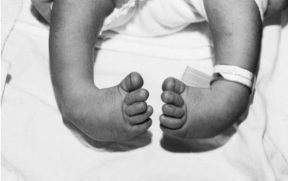
❓ Q1. Describe the findings in the image. What is the most likely diagnosis? List the four components of the deformity .
✅ Model Answer:
• Findings: Bilateral foot deformities – feet turned inward and downward. Heels in varus (inverted), forefoot adducted, ankle in equinus (plantarflexed). Rigid, not correctable to neutral.
• Diagnosis: Bilateral talipes equinovarus (congenital clubfoot).
• Four components (CAVE): Cavus (high arch), Adductus (forefoot adduction), Varus (heel inversion), Equinus (ankle plantarflexion).
• Findings: Bilateral foot deformities – feet turned inward and downward. Heels in varus (inverted), forefoot adducted, ankle in equinus (plantarflexed). Rigid, not correctable to neutral.
• Diagnosis: Bilateral talipes equinovarus (congenital clubfoot).
• Four components (CAVE): Cavus (high arch), Adductus (forefoot adduction), Varus (heel inversion), Equinus (ankle plantarflexion).
❓ Q2. What is the incidence of clubfoot? Is it more common in males or females? What is the etiology?
✅ Model Answer:
• Incidence: 1-2 per 1,000 live births. Male:Female = 2:1. Bilateral in ~50%.
• Etiology: Multifactorial (genetic + environmental). Strong genetic component (25% risk in first-degree relatives). Candidate genes: PITX1, TBX4, HOX.
• Associated conditions: Idiopathic (80%) or syndromic (20%) – myelomeningocele, arthrogryposis, amniotic band, trisomy 18/21.
• Environmental: Maternal smoking increases risk (2-3x).
• Incidence: 1-2 per 1,000 live births. Male:Female = 2:1. Bilateral in ~50%.
• Etiology: Multifactorial (genetic + environmental). Strong genetic component (25% risk in first-degree relatives). Candidate genes: PITX1, TBX4, HOX.
• Associated conditions: Idiopathic (80%) or syndromic (20%) – myelomeningocele, arthrogryposis, amniotic band, trisomy 18/21.
• Environmental: Maternal smoking increases risk (2-3x).
❓ Q3. What is the difference between structural (true) clubfoot and positional clubfoot (metatarsus adductus)?
✅ Model Answer:
• Structural clubfoot: Rigid deformity (cavus, adductus, varus, equinus). Cannot passively correct to neutral. Requires casting or surgery.
• Positional clubfoot (metatarsus adductus): Flexible, correctable to neutral. Only forefoot adductus, no cavus/varus/equinus. Resolves spontaneously with stretching or short-term casting.
• Other differentials: Calcaneovalgus foot (flexible, dorsiflexed), vertical talus (rigid rocker-bottom foot).
• Structural clubfoot: Rigid deformity (cavus, adductus, varus, equinus). Cannot passively correct to neutral. Requires casting or surgery.
• Positional clubfoot (metatarsus adductus): Flexible, correctable to neutral. Only forefoot adductus, no cavus/varus/equinus. Resolves spontaneously with stretching or short-term casting.
• Other differentials: Calcaneovalgus foot (flexible, dorsiflexed), vertical talus (rigid rocker-bottom foot).
❓ Q4. Describe the Ponseti method for treating clubfoot. What are the key steps?
✅ Model Answer:
• Gold standard non-surgical treatment, >90% success.
• Key steps: 1) Manipulation and serial casting (weekly, 5-8 casts) – correct cavus → adductus → varus → equinus. 2) Percutaneous Achilles tenotomy (90% of patients) to correct residual equinus. 3) Foot abduction orthosis (Denis Browne bar) – 23h/day for 3 months, then nighttime use until age 4-5 years.
• Start within 1-2 weeks of birth.
• Gold standard non-surgical treatment, >90% success.
• Key steps: 1) Manipulation and serial casting (weekly, 5-8 casts) – correct cavus → adductus → varus → equinus. 2) Percutaneous Achilles tenotomy (90% of patients) to correct residual equinus. 3) Foot abduction orthosis (Denis Browne bar) – 23h/day for 3 months, then nighttime use until age 4-5 years.
• Start within 1-2 weeks of birth.
❓ Q5. What is the percutaneous Achilles tenotomy? When is it performed? What are the risks?
✅ Model Answer:
• Definition: Minor surgical release of tight Achilles tendon to correct equinus.
• Timing: After 5-8 weekly casts (usually 6-8 weeks of age) when cavus/adductus/varus corrected but residual equinus remains.
• Technique: Under local anesthesia, small blade inserted percutaneously to divide tendon (felt as a pop). Final cast for 3 weeks.
• Risks: Bleeding, infection, sural nerve injury (rare), incomplete release. Very safe overall.
• After tenotomy, dorsiflexion improves from -20° to +15-20°.
• Definition: Minor surgical release of tight Achilles tendon to correct equinus.
• Timing: After 5-8 weekly casts (usually 6-8 weeks of age) when cavus/adductus/varus corrected but residual equinus remains.
• Technique: Under local anesthesia, small blade inserted percutaneously to divide tendon (felt as a pop). Final cast for 3 weeks.
• Risks: Bleeding, infection, sural nerve injury (rare), incomplete release. Very safe overall.
• After tenotomy, dorsiflexion improves from -20° to +15-20°.
❓ Q6. What is the foot abduction orthosis (Denis Browne bar)? How long should it be worn to prevent relapse?
✅ Model Answer:
• FAO (Denis Browne bar): Bar connecting two high-top boots holding feet in external rotation (70°) and dorsiflexion (15-20°). Prevents recurrence of varus/adductus.
• Wearing schedule: First 3 months – 23 hours/day (full-time). Next 2-4 years – nighttime and nap use only (12-14 hours/day). Total duration until 4-5 years of age.
• Compliance is the most important factor preventing relapse. Non-compliance → recurrence in up to 50%.
• FAO (Denis Browne bar): Bar connecting two high-top boots holding feet in external rotation (70°) and dorsiflexion (15-20°). Prevents recurrence of varus/adductus.
• Wearing schedule: First 3 months – 23 hours/day (full-time). Next 2-4 years – nighttime and nap use only (12-14 hours/day). Total duration until 4-5 years of age.
• Compliance is the most important factor preventing relapse. Non-compliance → recurrence in up to 50%.
❓ Q7. What is the rate of relapse after Ponseti treatment? How is relapse managed?
✅ Model Answer:
• Relapse rate: 10-30% (higher with bracing non-compliance, syndromic clubfoot, late treatment).
• Management: Mild relapse (<1 year) → repeat Ponseti casting (3-5 casts) ± repeat tenotomy. Moderate relapse (1-2 years) → casting ± tenotomy, strict bracing. Severe/late relapse (>2 years) or failed non-surgical → surgical release (posteromedial release, tendon transfer, osteotomy).
• Address bracing compliance.
• Relapse rate: 10-30% (higher with bracing non-compliance, syndromic clubfoot, late treatment).
• Management: Mild relapse (<1 year) → repeat Ponseti casting (3-5 casts) ± repeat tenotomy. Moderate relapse (1-2 years) → casting ± tenotomy, strict bracing. Severe/late relapse (>2 years) or failed non-surgical → surgical release (posteromedial release, tendon transfer, osteotomy).
• Address bracing compliance.
❓ Q8. What specific physical examination findings confirm the diagnosis of clubfoot? How do you assess severity?
✅ Model Answer:
• Inspection: Small, short foot with deep medial skin creases, empty heel, prominent talar head (palpable laterally). Cavus, adductus, varus, equinus.
• Mobility: Rigid – cannot passively bring foot to neutral.
• Severity scoring: Pirani score (6-point scale) – assesses posterior crease, empty heel, lateral border convex, medial crease, coverage of lateral head of talus, rigid equinus. Higher score = more severe.
• Neurological exam: Check for associated myelomeningocele.
• Inspection: Small, short foot with deep medial skin creases, empty heel, prominent talar head (palpable laterally). Cavus, adductus, varus, equinus.
• Mobility: Rigid – cannot passively bring foot to neutral.
• Severity scoring: Pirani score (6-point scale) – assesses posterior crease, empty heel, lateral border convex, medial crease, coverage of lateral head of talus, rigid equinus. Higher score = more severe.
• Neurological exam: Check for associated myelomeningocele.
❓ Q9. Which conditions are associated with clubfoot? When should you suspect a syndromic cause?
✅ Model Answer:
• Idiopathic clubfoot (80%) – isolated.
• Syndromic/associated (20%): Myelomeningocele (neurogenic), arthrogryposis multiplex congenita, amniotic band sequence, trisomy 18/21 (Down syndrome), neuromuscular disorders (SMA, CP), skeletal dysplasias (diastrophic dysplasia).
• Suspect syndromic cause if: Other anomalies (sacral dimple, hair tuft, dysmorphic facies, cardiac murmur, hypotonia), or clubfoot very rigid and resistant to casting.
• Idiopathic clubfoot (80%) – isolated.
• Syndromic/associated (20%): Myelomeningocele (neurogenic), arthrogryposis multiplex congenita, amniotic band sequence, trisomy 18/21 (Down syndrome), neuromuscular disorders (SMA, CP), skeletal dysplasias (diastrophic dysplasia).
• Suspect syndromic cause if: Other anomalies (sacral dimple, hair tuft, dysmorphic facies, cardiac murmur, hypotonia), or clubfoot very rigid and resistant to casting.
❓ Q10. What is the long-term prognosis for a child with idiopathic clubfoot treated with the Ponseti method?
✅ Model Answer:
• Excellent prognosis. >90% achieve functional, plantigrade foot with good mobility and no pain.
• Most wear normal shoes and participate in sports. Some have mild calf atrophy and slightly smaller foot size (0.5-1 size difference).
• Subtalar stiffness may persist but rarely limits function.
• Small risk of late relapse (up to 20%) – requires repeat casting or surgical revision.
• With Ponseti, <10% need extensive surgery (vs. 90% pre-Ponseti era).
• Excellent prognosis. >90% achieve functional, plantigrade foot with good mobility and no pain.
• Most wear normal shoes and participate in sports. Some have mild calf atrophy and slightly smaller foot size (0.5-1 size difference).
• Subtalar stiffness may persist but rarely limits function.
• Small risk of late relapse (up to 20%) – requires repeat casting or surgical revision.
• With Ponseti, <10% need extensive surgery (vs. 90% pre-Ponseti era).
❓ Q11. How will you counsel the parents of this newborn with bilateral clubfoot?
✅ Model Answer:
• “Your baby’s feet are turned inward/downward due to clubfoot – a condition from early pregnancy.”
• “We start treatment next week with the Ponseti method – gentle manipulation and weekly casts for 6-8 weeks. The baby will not be in pain.”
• “Most babies need a small Achilles tenotomy at the end of casting to allow full ankle motion – done under local anesthesia.”
• “After casting, your baby must wear boots attached to a bar (brace) for 3 months full-time, then at night until age 4-5 years. This is critical to prevent relapse.”
• “Success rate over 90% – most children have normal-looking feet and walk/run/play without limitations. This is not your fault.”
• “Your baby’s feet are turned inward/downward due to clubfoot – a condition from early pregnancy.”
• “We start treatment next week with the Ponseti method – gentle manipulation and weekly casts for 6-8 weeks. The baby will not be in pain.”
• “Most babies need a small Achilles tenotomy at the end of casting to allow full ankle motion – done under local anesthesia.”
• “After casting, your baby must wear boots attached to a bar (brace) for 3 months full-time, then at night until age 4-5 years. This is critical to prevent relapse.”
• “Success rate over 90% – most children have normal-looking feet and walk/run/play without limitations. This is not your fault.”
❓ Q12. Can clubfoot be diagnosed prenatally? What is the management if diagnosed in utero?
✅ Model Answer:
• Yes, diagnosed prenatally by ultrasound at 18-22 weeks (PPV 50-80%).
• Findings: Foot in persistent equinus/varus, inability to see normal foot alignment.
• Management: Detailed fetal anatomy scan (rule out spine, neural tube, chromosomal anomalies). Amniocentesis if other anomalies. Fetal echocardiogram if syndromic suspicion.
• Refer to pediatric orthopedist and neonatology. Reassure parents of excellent prognosis.
• Yes, diagnosed prenatally by ultrasound at 18-22 weeks (PPV 50-80%).
• Findings: Foot in persistent equinus/varus, inability to see normal foot alignment.
• Management: Detailed fetal anatomy scan (rule out spine, neural tube, chromosomal anomalies). Amniocentesis if other anomalies. Fetal echocardiogram if syndromic suspicion.
• Refer to pediatric orthopedist and neonatology. Reassure parents of excellent prognosis.
📋 Mock Test Feedback
💡 Examiner's note: Compare your answers with the model answers. In real TOACS, you would discuss these with the examiner.