⚕️ FCPS Paediatrics TOACS · Mock Test
| Observed Station | CPSP Format
⏱️ TIME REMAINING
08:00
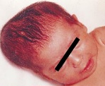
❓ Q1. Describe the findings in the image. What is the most likely diagnosis?
✅ Model Answer:
• Findings: Well-defined, firm, fluctuant, non-pulsatile swelling over the right parietal bone. Does NOT cross the sagittal suture line. Overlying skin normal, no erythema or abrasion.
• Diagnosis: Cephalohematoma – a subperiosteal hemorrhage typically associated with vacuum-assisted or forceps delivery.
• Findings: Well-defined, firm, fluctuant, non-pulsatile swelling over the right parietal bone. Does NOT cross the sagittal suture line. Overlying skin normal, no erythema or abrasion.
• Diagnosis: Cephalohematoma – a subperiosteal hemorrhage typically associated with vacuum-assisted or forceps delivery.
❓ Q2. What is the underlying pathology of cephalohematoma? Why does it not cross suture lines?
✅ Model Answer:
• Pathology: Bleeding into the subperiosteal space between skull periosteum and calvarial bone due to rupture of small emissary/diploic veins during birth trauma.
• The periosteum is tightly adherent to the skull bone at suture lines (sutures are periosteal reflections). Therefore hematoma is confined to a single cranial bone and does NOT cross sutures.
• Risk factors: Vacuum extraction (most common), forceps delivery, prolonged labor, macrosomia, primiparity.
• Incidence: 1-2% of live births; up to 10-15% with instrumental delivery.
• Pathology: Bleeding into the subperiosteal space between skull periosteum and calvarial bone due to rupture of small emissary/diploic veins during birth trauma.
• The periosteum is tightly adherent to the skull bone at suture lines (sutures are periosteal reflections). Therefore hematoma is confined to a single cranial bone and does NOT cross sutures.
• Risk factors: Vacuum extraction (most common), forceps delivery, prolonged labor, macrosomia, primiparity.
• Incidence: 1-2% of live births; up to 10-15% with instrumental delivery.
❓ Q3. What are the differential diagnoses of a neonatal scalp swelling? Differentiate from caput succedaneum and subgaleal hemorrhage.
✅ Model Answer:
• Cephalohematoma: Subperiosteal, confined to one bone, does not cross sutures, firm/fluctuant, appears hours to days after birth, resolves over weeks to months.
• Caput succedaneum: Subcutaneous edema, crosses suture lines, soft/pitting, present at birth, resolves within days (24-72 hours).
• Subgaleal hemorrhage: Subaponeurotic (potential space), crosses sutures, fluctuant (can spread to entire scalp), massive (up to 40% blood volume), hypovolemic shock, emergency. Associated with vacuum extraction.
• Cephalohematoma: Subperiosteal, confined to one bone, does not cross sutures, firm/fluctuant, appears hours to days after birth, resolves over weeks to months.
• Caput succedaneum: Subcutaneous edema, crosses suture lines, soft/pitting, present at birth, resolves within days (24-72 hours).
• Subgaleal hemorrhage: Subaponeurotic (potential space), crosses sutures, fluctuant (can spread to entire scalp), massive (up to 40% blood volume), hypovolemic shock, emergency. Associated with vacuum extraction.
❓ Q4. What is the natural history of cephalohematoma? How long does it take to resolve?
✅ Model Answer:
• Spontaneous resolution in most cases without intervention.
• Timeline: Swelling may increase over first 24-48 hours → firmness increases over first week → gradual resorption begins by 2-3 weeks → complete resolution typically by 2-4 months (up to 6 months).
• May leave a calcified rim (bony ridge) that resolves over months to years.
• No treatment needed. Avoid aspiration – risk of infection (osteomyelitis).
• Spontaneous resolution in most cases without intervention.
• Timeline: Swelling may increase over first 24-48 hours → firmness increases over first week → gradual resorption begins by 2-3 weeks → complete resolution typically by 2-4 months (up to 6 months).
• May leave a calcified rim (bony ridge) that resolves over months to years.
• No treatment needed. Avoid aspiration – risk of infection (osteomyelitis).
❓ Q5. What are the potential complications of cephalohematoma?
✅ Model Answer:
• Hyperbilirubinemia (most common, 20-30%) – breakdown of hematoma releases hemoglobin → increased bilirubin load.
• Anemia (rare, large hematomas) – blood sequestration up to 50-100 mL.
• Infection/osteomyelitis (very rare) – if aspiration attempted. Presents with erythema, warmth, tenderness, fever.
• Associated linear skull fracture (10-25%) – usually incidental, no treatment.
• Calcification/ossification – may leave a "bony knob".
• Generally painless; infant usually asymptomatic.
• Hyperbilirubinemia (most common, 20-30%) – breakdown of hematoma releases hemoglobin → increased bilirubin load.
• Anemia (rare, large hematomas) – blood sequestration up to 50-100 mL.
• Infection/osteomyelitis (very rare) – if aspiration attempted. Presents with erythema, warmth, tenderness, fever.
• Associated linear skull fracture (10-25%) – usually incidental, no treatment.
• Calcification/ossification – may leave a "bony knob".
• Generally painless; infant usually asymptomatic.
❓ Q6. How does cephalohematoma contribute to hyperbilirubinemia? How do you monitor and manage it?
✅ Model Answer:
• Mechanism: Breakdown of extravasated RBCs increases bilirubin load on neonatal liver (heme oxygenase pathway). Immature liver cannot conjugate increased bilirubin → unconjugated hyperbilirubinemia.
• Monitoring: Serial bilirubin measurements (transcutaneous or serum) every 12-24 hours for first 3-5 days, especially large hematoma. Monitor jaundice progression.
• Management: Ensure adequate hydration/frequent feeds. Phototherapy if bilirubin exceeds age-specific nomogram thresholds. Exchange transfusion rarely needed.
• Mechanism: Breakdown of extravasated RBCs increases bilirubin load on neonatal liver (heme oxygenase pathway). Immature liver cannot conjugate increased bilirubin → unconjugated hyperbilirubinemia.
• Monitoring: Serial bilirubin measurements (transcutaneous or serum) every 12-24 hours for first 3-5 days, especially large hematoma. Monitor jaundice progression.
• Management: Ensure adequate hydration/frequent feeds. Phototherapy if bilirubin exceeds age-specific nomogram thresholds. Exchange transfusion rarely needed.
❓ Q7. Can a cephalohematoma cause significant anemia? When should you suspect it?
✅ Model Answer:
• Yes, but rare. Very large cephalohematomas can sequester 50-100 mL of blood (neonatal blood volume ~80-100 mL/kg).
• Suspect anemia if: Pale appearance, poor feeding, lethargy, tachycardia, tachypnea; hematoma >6-8 cm diameter; fall in hemoglobin/hematocrit on serial CBC.
• Management: Monitor hemoglobin. If symptomatic and Hb <10 g/dL → packed RBC transfusion. Iron supplementation after resolution.
• Yes, but rare. Very large cephalohematomas can sequester 50-100 mL of blood (neonatal blood volume ~80-100 mL/kg).
• Suspect anemia if: Pale appearance, poor feeding, lethargy, tachycardia, tachypnea; hematoma >6-8 cm diameter; fall in hemoglobin/hematocrit on serial CBC.
• Management: Monitor hemoglobin. If symptomatic and Hb <10 g/dL → packed RBC transfusion. Iron supplementation after resolution.
❓ Q8. What is the risk of osteomyelitis with cephalohematoma? How does it present and how is it treated?
✅ Model Answer:
• Risk: Extremely low (<1%) unless aspiration or needle drainage attempted.
• Presentation (2-6 weeks): Swelling becomes erythematous, warm, tender; fever, irritability, elevated inflammatory markers; associated skull osteomyelitis.
• Pathogens: S. aureus, Group B Strep, E. coli.
• Management: IV antibiotics (vancomycin + cefotaxime or cloxacillin + gentamicin). Imaging (US or MRI). Surgical drainage if abscess. Antibiotics 3-6 weeks.
• Prevention: Do NOT aspirate or drain uninfected cephalohematoma.
• Risk: Extremely low (<1%) unless aspiration or needle drainage attempted.
• Presentation (2-6 weeks): Swelling becomes erythematous, warm, tender; fever, irritability, elevated inflammatory markers; associated skull osteomyelitis.
• Pathogens: S. aureus, Group B Strep, E. coli.
• Management: IV antibiotics (vancomycin + cefotaxime or cloxacillin + gentamicin). Imaging (US or MRI). Surgical drainage if abscess. Antibiotics 3-6 weeks.
• Prevention: Do NOT aspirate or drain uninfected cephalohematoma.
❓ Q9. When is imaging (skull X-ray, CT, ultrasound) indicated for a cephalohematoma?
✅ Model Answer:
• Routine imaging NOT indicated for typical uncomplicated cephalohematoma – diagnosis is clinical.
• Indications for skull X-ray: Suspected underlying depressed skull fracture, neurological symptoms (seizures, altered sensorium), large/expanding hematoma beyond 48 hours.
• Indications for ultrasound/CT: Suspected subgaleal hemorrhage (crosses sutures rapidly), suspected infection (abscess, osteomyelitis), neurological symptoms.
• Finding: Skull X-ray may show linear skull fracture in 10-25% – usually no intervention.
• Routine imaging NOT indicated for typical uncomplicated cephalohematoma – diagnosis is clinical.
• Indications for skull X-ray: Suspected underlying depressed skull fracture, neurological symptoms (seizures, altered sensorium), large/expanding hematoma beyond 48 hours.
• Indications for ultrasound/CT: Suspected subgaleal hemorrhage (crosses sutures rapidly), suspected infection (abscess, osteomyelitis), neurological symptoms.
• Finding: Skull X-ray may show linear skull fracture in 10-25% – usually no intervention.
❓ Q10. What is the association between cephalohematoma and skull fracture? How is it managed?
✅ Model Answer:
• Incidence: Underlying linear skull fracture in 10-25% of cephalohematomas (especially after forceps delivery).
• Linear non-depressed fractures require no treatment – heal spontaneously in weeks to months.
• Depressed ("ping-pong") fractures: Rare; may require surgical elevation if neurological deficit or cosmetic concerns.
• Monitor for complications: leptomeningeal cyst (growing skull fracture) – extremely rare.
• Do NOT routinely image every cephalohematoma; only if neurological signs or depressed fracture palpable.
• Incidence: Underlying linear skull fracture in 10-25% of cephalohematomas (especially after forceps delivery).
• Linear non-depressed fractures require no treatment – heal spontaneously in weeks to months.
• Depressed ("ping-pong") fractures: Rare; may require surgical elevation if neurological deficit or cosmetic concerns.
• Monitor for complications: leptomeningeal cyst (growing skull fracture) – extremely rare.
• Do NOT routinely image every cephalohematoma; only if neurological signs or depressed fracture palpable.
❓ Q11. How will you manage this infant? What will you tell the anxious mother?
✅ Model Answer:
• Management: Observation, NO intervention.
• “This is a cephalohematoma – a collection of blood under the outer covering of the skull bone. It is NOT inside the brain and does NOT affect the brain.”
• “It will go away completely on its own over 2-4 months. No treatment or medicine is needed.”
• “Do NOT apply anything to it, do NOT massage it, and do NOT try to drain it – squeezing can cause serious infection.”
• “We will monitor your baby for jaundice (yellow skin) from breakdown of blood.”
• “If the swelling becomes red, warm, tender, or your baby develops fever – come back immediately.”
• Management: Observation, NO intervention.
• “This is a cephalohematoma – a collection of blood under the outer covering of the skull bone. It is NOT inside the brain and does NOT affect the brain.”
• “It will go away completely on its own over 2-4 months. No treatment or medicine is needed.”
• “Do NOT apply anything to it, do NOT massage it, and do NOT try to drain it – squeezing can cause serious infection.”
• “We will monitor your baby for jaundice (yellow skin) from breakdown of blood.”
• “If the swelling becomes red, warm, tender, or your baby develops fever – come back immediately.”
❓ Q12. What follow-up is required? When would you refer to a neurosurgeon or pediatric surgeon?
✅ Model Answer:
• Follow-up: Routine newborn care; re-examine at 2-week and 2-month visits. Monitor bilirubin for first 5 days if large hematoma. Document size and resolution.
• Referral indications: Depressed skull fracture with neurological deficit, growing skull fracture (leptomeningeal cyst), suspected osteomyelitis/abscess, cosmetically concerning persistent calcified ridge after 1 year (rare).
• Most cephalohematomas do NOT require subspecialty referral.
• Follow-up: Routine newborn care; re-examine at 2-week and 2-month visits. Monitor bilirubin for first 5 days if large hematoma. Document size and resolution.
• Referral indications: Depressed skull fracture with neurological deficit, growing skull fracture (leptomeningeal cyst), suspected osteomyelitis/abscess, cosmetically concerning persistent calcified ridge after 1 year (rare).
• Most cephalohematomas do NOT require subspecialty referral.
📋 Mock Test Feedback
💡 Examiner's note: Compare your answers with the model answers. In real TOACS, you would discuss these with the examiner.