FCPS MCPS IMM MD Paediatrics TOACS
Observed Station | CPSP Format | 8 minutes
⏱️ TIME REMAINING
08:00
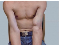
A 9‑year‑old child is brought in due to a rapidly enlarging mass on the left arm, associated with severe pain and limited movement,The child has multiple cafe‑au‑lait spots.
❓ Q1. Identify the lesion shown in the image. What condition is it associated with?
✅ Model Answer: • Lesion: Plexiform neurofibroma – a diffuse, infiltrating, benign peripheral nerve sheath tumor.
• Associated condition: Neurofibromatosis type 1 (NF1).
• Key features: Congenital or early childhood onset, soft "bag of worms" texture, overlying hyperpigmented skin, can involve any nerve (commonly cervical, facial, lumbosacral).
• Associated condition: Neurofibromatosis type 1 (NF1).
• Key features: Congenital or early childhood onset, soft "bag of worms" texture, overlying hyperpigmented skin, can involve any nerve (commonly cervical, facial, lumbosacral).
❓ Q2. What is the genetic basis of neurofibromatosis type 1?
✅ Model Answer: • Gene: NF1 gene located on chromosome 17q11.2.
• Protein: Neurofibromin – a tumor suppressor protein that negatively regulates the Ras-MAPK signaling pathway (Ras GTPase-activating protein).
• Inheritance: Autosomal dominant (50% de novo mutations).
• Pathogenesis: Loss of neurofibromin → increased Ras signaling → uncontrolled cell proliferation and tumor formation.
• Protein: Neurofibromin – a tumor suppressor protein that negatively regulates the Ras-MAPK signaling pathway (Ras GTPase-activating protein).
• Inheritance: Autosomal dominant (50% de novo mutations).
• Pathogenesis: Loss of neurofibromin → increased Ras signaling → uncontrolled cell proliferation and tumor formation.
❓ Q3. What are the NIH diagnostic criteria for neurofibromatosis type 1 (NF1)?
✅ Model Answer: • NIH diagnostic criteria (≥2 of the following):
1. Café-au-lait macules: ≥6 of >0.5 cm (prepubertal) or >1.5 cm (postpubertal).
2. Axillary or inguinal freckling.
3. Neurofibromas: ≥2 cutaneous neurofibromas OR ≥1 plexiform neurofibroma.
4. Optic pathway glioma.
5. Lisch nodules (iris hamartomas) – ≥2 on slit-lamp exam.
6. Sphenoid wing dysplasia OR tibial pseudarthrosis.
7. First-degree relative with NF1.
• Note: Revised criteria add pathogenic NF1 variant as a criterion.
1. Café-au-lait macules: ≥6 of >0.5 cm (prepubertal) or >1.5 cm (postpubertal).
2. Axillary or inguinal freckling.
3. Neurofibromas: ≥2 cutaneous neurofibromas OR ≥1 plexiform neurofibroma.
4. Optic pathway glioma.
5. Lisch nodules (iris hamartomas) – ≥2 on slit-lamp exam.
6. Sphenoid wing dysplasia OR tibial pseudarthrosis.
7. First-degree relative with NF1.
• Note: Revised criteria add pathogenic NF1 variant as a criterion.
❓ Q4. What are the clinical features of a plexiform neurofibroma?
✅ Model Answer: • Clinical features:
- Appearance: Diffuse, infiltrating, soft, "bag of worms" texture (palpable along nerve bundles).
- Onset: Usually congenital or presents in early childhood.
- Location: Head/neck (20%), trunk, extremities, lumbosacral, mediastinal.
- Overlying skin: Often hyperpigmented (café-au-lait spots).
- Associated features: May cause pain, disfigurement, functional impairment (e.g., limited movement, vision loss if periorbital).
- Growth: Can grow rapidly, especially during puberty or pregnancy.
- Appearance: Diffuse, infiltrating, soft, "bag of worms" texture (palpable along nerve bundles).
- Onset: Usually congenital or presents in early childhood.
- Location: Head/neck (20%), trunk, extremities, lumbosacral, mediastinal.
- Overlying skin: Often hyperpigmented (café-au-lait spots).
- Associated features: May cause pain, disfigurement, functional impairment (e.g., limited movement, vision loss if periorbital).
- Growth: Can grow rapidly, especially during puberty or pregnancy.
❓ Q5. What is the risk of malignant transformation of a plexiform neurofibroma?
✅ Model Answer: • Risk: 8-13% lifetime risk of malignant transformation to malignant peripheral nerve sheath tumor (MPNST).
• MPNST is the most common cause of death in NF1 patients.
• Red flags for transformation:
- Rapid growth (increase in size over weeks to months).
- New or worsening persistent pain (especially at rest).
- Neurologic deficit (weakness, numbness, or loss of function).
- Change in texture (hardening or firmness).
- PET/CT: SUVmax >3 is suggestive of malignancy.
• MPNST is the most common cause of death in NF1 patients.
• Red flags for transformation:
- Rapid growth (increase in size over weeks to months).
- New or worsening persistent pain (especially at rest).
- Neurologic deficit (weakness, numbness, or loss of function).
- Change in texture (hardening or firmness).
- PET/CT: SUVmax >3 is suggestive of malignancy.
❓ Q6. What imaging studies are useful for evaluating a plexiform neurofibroma?
✅ Model Answer: • MRI (with contrast): The imaging modality of choice. Shows the extent and infiltration of the lesion, which often has a characteristic "target sign" (central T2 hyperintensity).
• PET/CT (FDG-PET): Used to assess for malignant transformation. SUVmax >3 suggests MPNST; SUVmax >6 is highly suspicious. Biopsy should be considered if SUVmax is elevated.
• Ultrasound: May be used to assess vascularity and guide biopsy.
• Routine surveillance: MRI is not routinely performed for asymptomatic stable lesions; reserved for symptomatic or rapidly growing lesions.
• PET/CT (FDG-PET): Used to assess for malignant transformation. SUVmax >3 suggests MPNST; SUVmax >6 is highly suspicious. Biopsy should be considered if SUVmax is elevated.
• Ultrasound: May be used to assess vascularity and guide biopsy.
• Routine surveillance: MRI is not routinely performed for asymptomatic stable lesions; reserved for symptomatic or rapidly growing lesions.
❓ Q7. What is the role of selumetinib in the treatment of plexiform neurofibromas?
✅ Model Answer: • Selumetinib: A MEK1/MEK2 inhibitor (targeting the MAPK pathway).
• FDA approval: Approved in 2020 for pediatric patients (≥2 years) with inoperable, symptomatic plexiform neurofibromas in NF1.
• Mechanism: Blocks the downstream signaling of Ras-MAPK, which is hyperactivated due to neurofibromin deficiency.
• Benefits: Reduces tumor volume, improves pain and functional impairment.
• Dose: 25 mg/m² twice daily (oral).
• Side effects: GI symptoms (diarrhea, nausea), rash, fatigue, ocular toxicity (retinal pigment epithelial detachment – monitor with eye exams).
• FDA approval: Approved in 2020 for pediatric patients (≥2 years) with inoperable, symptomatic plexiform neurofibromas in NF1.
• Mechanism: Blocks the downstream signaling of Ras-MAPK, which is hyperactivated due to neurofibromin deficiency.
• Benefits: Reduces tumor volume, improves pain and functional impairment.
• Dose: 25 mg/m² twice daily (oral).
• Side effects: GI symptoms (diarrhea, nausea), rash, fatigue, ocular toxicity (retinal pigment epithelial detachment – monitor with eye exams).
❓ Q8. What is the role of surgical resection in plexiform neurofibromas?
✅ Model Answer: • Surgical role:
- Complete resection: The goal, but often not possible due to infiltration of surrounding tissues and nerves. Complete resection may be achievable for smaller, well-circumscribed lesions.
- Debulking: Performed for symptomatic relief (pain, disfigurement, functional impairment) or if malignancy is suspected (biopsy or excision).
- Challenges: High risk of nerve damage, recurrence, and bleeding. Incomplete resection is common.
- Indications for surgery: Rapid growth, severe pain, neurologic deficit, cosmetic concerns, or suspected MPNST.
- Multidisciplinary approach: Requires input from neurosurgery, orthopedics, and plastic surgery.
- Complete resection: The goal, but often not possible due to infiltration of surrounding tissues and nerves. Complete resection may be achievable for smaller, well-circumscribed lesions.
- Debulking: Performed for symptomatic relief (pain, disfigurement, functional impairment) or if malignancy is suspected (biopsy or excision).
- Challenges: High risk of nerve damage, recurrence, and bleeding. Incomplete resection is common.
- Indications for surgery: Rapid growth, severe pain, neurologic deficit, cosmetic concerns, or suspected MPNST.
- Multidisciplinary approach: Requires input from neurosurgery, orthopedics, and plastic surgery.
❓ Q9. What is the difference between a plexiform neurofibroma and a cutaneous neurofibroma?
✅ Model Answer: • Plexiform neurofibroma:
- Diffuse, infiltrating mass involving multiple nerve fascicles.
- Congenital or early childhood onset.
- May be large, disfiguring, and cause functional impairment.
- Risk of malignant transformation (MPNST).
- Diagnostic criterion for NF1 (≥1 plexiform neurofibroma).
• Cutaneous neurofibroma:
- Discrete, well-circumscribed, pedunculated or sessile lesions on the skin.
- Usually appear in adolescence or adulthood.
- Do NOT undergo malignant transformation.
- Not a diagnostic criterion for NF1 (≥2 cutaneous neurofibromas = 1 criterion).
- Can be numerous and cause cosmetic concerns.
- Diffuse, infiltrating mass involving multiple nerve fascicles.
- Congenital or early childhood onset.
- May be large, disfiguring, and cause functional impairment.
- Risk of malignant transformation (MPNST).
- Diagnostic criterion for NF1 (≥1 plexiform neurofibroma).
• Cutaneous neurofibroma:
- Discrete, well-circumscribed, pedunculated or sessile lesions on the skin.
- Usually appear in adolescence or adulthood.
- Do NOT undergo malignant transformation.
- Not a diagnostic criterion for NF1 (≥2 cutaneous neurofibromas = 1 criterion).
- Can be numerous and cause cosmetic concerns.
❓ Q10. What are the other malignancies associated with NF1?
✅ Model Answer: • Malignancies in NF1:
- MPNST (malignant peripheral nerve sheath tumor): Most common (8-13% lifetime risk).
- Optic glioma: Low-grade pilocytic astrocytoma (can cause vision loss).
- Other gliomas: Brainstem glioma, astrocytoma (other locations).
- Leukemia: Juvenile myelomonocytic leukemia (JMML) – especially in children <5 years.
- Breast cancer: Increased risk in adults with NF1 (recommended screening).
- Gastrointestinal stromal tumors (GISTs).
- Neuroblastoma (rare).
- Rhabdomyosarcoma (rare).
- MPNST (malignant peripheral nerve sheath tumor): Most common (8-13% lifetime risk).
- Optic glioma: Low-grade pilocytic astrocytoma (can cause vision loss).
- Other gliomas: Brainstem glioma, astrocytoma (other locations).
- Leukemia: Juvenile myelomonocytic leukemia (JMML) – especially in children <5 years.
- Breast cancer: Increased risk in adults with NF1 (recommended screening).
- Gastrointestinal stromal tumors (GISTs).
- Neuroblastoma (rare).
- Rhabdomyosarcoma (rare).
❓ Q11. What is the role of surveillance in NF1?
✅ Model Answer: • Surveillance recommendations:
- Ophthalmology: Annual eye exams (slit-lamp) for Lisch nodules and optic glioma (especially in children <8 years).
- Blood pressure: Monitor annually (risk of renovascular hypertension due to renal artery stenosis).
- Developmental screening: Assess for learning disabilities, ADHD, and cognitive deficits (common in NF1).
- Scoliosis screening: Annual physical exam.
- Skin: Annual skin exam for new/changing lesions.
- Neurologic: Neurological exam for any new deficits.
- Imaging: MRI for new or changing symptoms (not routine for stable lesions).
- Genetic counseling: For families; discuss recurrence risk and prenatal testing.
- Ophthalmology: Annual eye exams (slit-lamp) for Lisch nodules and optic glioma (especially in children <8 years).
- Blood pressure: Monitor annually (risk of renovascular hypertension due to renal artery stenosis).
- Developmental screening: Assess for learning disabilities, ADHD, and cognitive deficits (common in NF1).
- Scoliosis screening: Annual physical exam.
- Skin: Annual skin exam for new/changing lesions.
- Neurologic: Neurological exam for any new deficits.
- Imaging: MRI for new or changing symptoms (not routine for stable lesions).
- Genetic counseling: For families; discuss recurrence risk and prenatal testing.
❓ Q12. What is the differential diagnosis of a plexiform neurofibroma?
✅ Model Answer: • Differential diagnoses:
- MPNST (malignant peripheral nerve sheath tumor): Rapid growth, pain, neurologic deficit; requires biopsy to differentiate.
- Neurofibromatosis type 2 (NF2): Bilateral vestibular schwannomas; not associated with plexiform neurofibromas.
- Schwannoma: Well-circumscribed, slow-growing, non-infiltrating.
- Lipoma: Soft, fatty tumor; well-circumscribed, not infiltrating.
- Hemangioma: Vascular lesion; may be soft and compressible.
- Lymphatic malformation: Soft, cystic, often transilluminating.
- Rhabdomyosarcoma: Malignant soft tissue tumor; can present as a rapidly growing mass.
- MPNST (malignant peripheral nerve sheath tumor): Rapid growth, pain, neurologic deficit; requires biopsy to differentiate.
- Neurofibromatosis type 2 (NF2): Bilateral vestibular schwannomas; not associated with plexiform neurofibromas.
- Schwannoma: Well-circumscribed, slow-growing, non-infiltrating.
- Lipoma: Soft, fatty tumor; well-circumscribed, not infiltrating.
- Hemangioma: Vascular lesion; may be soft and compressible.
- Lymphatic malformation: Soft, cystic, often transilluminating.
- Rhabdomyosarcoma: Malignant soft tissue tumor; can present as a rapidly growing mass.
❓ Q13. How would you counsel the parents of a child with NF1 and a plexiform neurofibroma?
✅ Model Answer: • "Your child has neurofibromatosis type 1 (NF1), a genetic condition that causes tumors to grow along nerves."
• "The large mass is called a plexiform neurofibroma. It is a benign (non-cancerous) tumor that grows along the nerve bundles. Many children with NF1 have these."
• "We will monitor this lesion closely. If it grows rapidly, becomes painful, or causes functional problems, we will investigate further and consider treatment."
• "There is a medication called selumetinib that can shrink these tumors and improve symptoms. It is used in children with inoperable plexiform neurofibromas."
• "Regular check-ups are important to monitor for changes, not just in this lesion but also for other features of NF1 like optic gliomas, high blood pressure, and learning issues."
• "We are here to support you and your child through this lifelong journey."
• "The large mass is called a plexiform neurofibroma. It is a benign (non-cancerous) tumor that grows along the nerve bundles. Many children with NF1 have these."
• "We will monitor this lesion closely. If it grows rapidly, becomes painful, or causes functional problems, we will investigate further and consider treatment."
• "There is a medication called selumetinib that can shrink these tumors and improve symptoms. It is used in children with inoperable plexiform neurofibromas."
• "Regular check-ups are important to monitor for changes, not just in this lesion but also for other features of NF1 like optic gliomas, high blood pressure, and learning issues."
• "We are here to support you and your child through this lifelong journey."
❓ Q14. What is the role of MPNST in NF1?
✅ Model Answer: • MPNST (malignant peripheral nerve sheath tumor):
- Incidence: 8-13% lifetime risk in NF1 patients.
- Most common cause of death in NF1.
- Origin: Usually arises from a pre-existing plexiform neurofibroma.
- Presentation: Rapid growth, persistent pain (especially at rest), neurologic deficit, and change in texture (hardening).
- Imaging: PET/CT – SUVmax >3 suggests malignancy.
- Treatment: Surgical resection (if feasible), chemotherapy (ifosfamide, doxorubicin), radiation therapy.
- Prognosis: Poor – 5-year survival <20% for metastatic MPNST.
- Incidence: 8-13% lifetime risk in NF1 patients.
- Most common cause of death in NF1.
- Origin: Usually arises from a pre-existing plexiform neurofibroma.
- Presentation: Rapid growth, persistent pain (especially at rest), neurologic deficit, and change in texture (hardening).
- Imaging: PET/CT – SUVmax >3 suggests malignancy.
- Treatment: Surgical resection (if feasible), chemotherapy (ifosfamide, doxorubicin), radiation therapy.
- Prognosis: Poor – 5-year survival <20% for metastatic MPNST.
❓ Q15. What is the role of genetic testing in NF1?
✅ Model Answer: • Role of genetic testing:
- Confirm diagnosis: Identifies pathogenic variants in the NF1 gene (detection rate ~95%).
- Differentiate from other conditions: NF2, Legius syndrome (SPRED1), schwannomatosis.
- Family counseling: Helps identify at-risk family members and discuss inheritance (autosomal dominant).
- Prenatal testing: For families with known mutations (CVS or amniocentesis).
- Clinical utility: Early diagnosis allows for appropriate surveillance and management.
- Note: Clinical diagnosis is sufficient in patients meeting NIH criteria; genetic testing is not required but can be helpful in ambiguous cases.
- Confirm diagnosis: Identifies pathogenic variants in the NF1 gene (detection rate ~95%).
- Differentiate from other conditions: NF2, Legius syndrome (SPRED1), schwannomatosis.
- Family counseling: Helps identify at-risk family members and discuss inheritance (autosomal dominant).
- Prenatal testing: For families with known mutations (CVS or amniocentesis).
- Clinical utility: Early diagnosis allows for appropriate surveillance and management.
- Note: Clinical diagnosis is sufficient in patients meeting NIH criteria; genetic testing is not required but can be helpful in ambiguous cases.
📋 Mock Test Feedback
💡 Examiner's note: Compare your answers with model answers. In real TOACS, you would discuss these with the examiner.