⚕️ FCPS MCPS IMM MD Paediatrics TOACS · Mock Test
X-ray Long Bones · 8-Minute Observed Station
📚 paeds.online
⏱️ TIME REMAINING
08:00
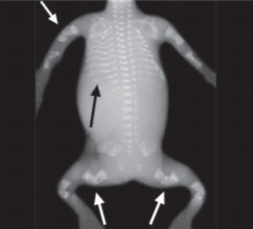
❓ Q1. Describe the radiographic findings. What is the most likely diagnosis?
✅ Model Answer:
• Multiple fractures – healing and healed fractures at various stages.
• Bowed long bones – femoral and tibial bowing (especially in Type III).
• Osteopenia – decreased bone density with thin cortices.
• Popcorn calcifications – metaphyseal areas (Type III).
• Diagnosis: Osteogenesis imperfecta (likely Type III or IV).
• Multiple fractures – healing and healed fractures at various stages.
• Bowed long bones – femoral and tibial bowing (especially in Type III).
• Osteopenia – decreased bone density with thin cortices.
• Popcorn calcifications – metaphyseal areas (Type III).
• Diagnosis: Osteogenesis imperfecta (likely Type III or IV).
❓ Q2. What are the key radiographic features of osteogenesis imperfecta on long bone X-rays?
✅ Model Answer:
• Multiple fractures – at various stages of healing (acute, healing, healed).
• Bowed long bones – especially femur and tibia (Type III).
• Osteopenia – decreased bone density, thin cortices, and trabecular rarefaction.
• Popcorn calcifications – nodular calcifications in the metaphyses (Type III).
• Thin cortices – with medullary expansion (due to multiple fractures).
• Wormian bones – on skull X-ray (intrasutural bones).
• "Bone-within-bone" appearance – may be seen in some cases.
• Multiple fractures – at various stages of healing (acute, healing, healed).
• Bowed long bones – especially femur and tibia (Type III).
• Osteopenia – decreased bone density, thin cortices, and trabecular rarefaction.
• Popcorn calcifications – nodular calcifications in the metaphyses (Type III).
• Thin cortices – with medullary expansion (due to multiple fractures).
• Wormian bones – on skull X-ray (intrasutural bones).
• "Bone-within-bone" appearance – may be seen in some cases.
❓ Q3. What is the pathophysiology of osteogenesis imperfecta?
✅ Model Answer:
• Osteogenesis imperfecta (OI) is a genetic disorder caused by mutations in the COL1A1 and COL1A2 genes (type I collagen).
• Type I collagen is the most abundant protein in bone, skin, tendons, ligaments, and dentin.
• Mutations:
- COL1A1 (chromosome 17) – more common in Type I.
- COL1A2 (chromosome 7) – less common.
• The mutations lead to reduced production or structural defects in type I collagen → bone fragility and reduced bone density.
• This results in brittle bones that fracture easily with minimal trauma.
• Inheritance: Autosomal dominant (most cases), autosomal recessive (rare).
• Other features: Blue sclerae (thinned sclera), dentinogenesis imperfecta, hearing loss.
• Osteogenesis imperfecta (OI) is a genetic disorder caused by mutations in the COL1A1 and COL1A2 genes (type I collagen).
• Type I collagen is the most abundant protein in bone, skin, tendons, ligaments, and dentin.
• Mutations:
- COL1A1 (chromosome 17) – more common in Type I.
- COL1A2 (chromosome 7) – less common.
• The mutations lead to reduced production or structural defects in type I collagen → bone fragility and reduced bone density.
• This results in brittle bones that fracture easily with minimal trauma.
• Inheritance: Autosomal dominant (most cases), autosomal recessive (rare).
• Other features: Blue sclerae (thinned sclera), dentinogenesis imperfecta, hearing loss.
❓ Q4. What is the Sillence classification of osteogenesis imperfecta?
✅ Model Answer:
| Type | Sclerae | Severity | Key Features |
|---|---|---|---|
| Type I | Blue | Mild | Fractures in childhood, hearing loss (20-30%), normal stature, dentinogenesis imperfecta (mild) |
| Type II | Blue | Lethal | Perinatal death, multiple fractures in utero, crumpled femurs, beaded ribs |
| Type III | White | Severe | Bowed femurs, progressive deformity, short stature, triangular facies, popcorn calcifications |
| Type IV | White | Moderate | Variable fractures, short stature, dentinogenesis imperfecta (common) |
❓ Q5. A child with OI Type III presents with bowed femurs and multiple fractures. What is the management?
✅ Model Answer:
• Medical management:
1. Bisphosphonates: IV pamidronate (or zoledronic acid) – increases bone density and reduces fracture risk. Protocol: 1 mg/kg/day IV for 3 consecutive days, repeated every 3-4 months.
2. Calcium and vitamin D supplementation: To support bone health.
3. Pain management: Analgesics for acute fractures.
• Surgical management:
1. Intramedullary rodding: Telescoping rods (Sofield-Millar) to straighten and stabilize bowed long bones.
2. Fracture fixation: For acute fractures.
• Rehabilitation:
1. Physical therapy: To maintain muscle strength and joint mobility.
2. Occupational therapy: For daily living activities.
3. Hydrotherapy: To improve mobility with low impact.
• Multidisciplinary care: Genetics, orthopedics, dentistry, audiology, and physiotherapy.
• Medical management:
1. Bisphosphonates: IV pamidronate (or zoledronic acid) – increases bone density and reduces fracture risk. Protocol: 1 mg/kg/day IV for 3 consecutive days, repeated every 3-4 months.
2. Calcium and vitamin D supplementation: To support bone health.
3. Pain management: Analgesics for acute fractures.
• Surgical management:
1. Intramedullary rodding: Telescoping rods (Sofield-Millar) to straighten and stabilize bowed long bones.
2. Fracture fixation: For acute fractures.
• Rehabilitation:
1. Physical therapy: To maintain muscle strength and joint mobility.
2. Occupational therapy: For daily living activities.
3. Hydrotherapy: To improve mobility with low impact.
• Multidisciplinary care: Genetics, orthopedics, dentistry, audiology, and physiotherapy.
❓ Q6. What is the role of bisphosphonates in osteogenesis imperfecta?
✅ Model Answer:
• Bisphosphonates (pamidronate, zoledronic acid) are the mainstay of medical therapy for OI.
• Mechanism: Inhibit osteoclast-mediated bone resorption → increase bone mineral density (BMD) and reduce fracture risk.
• Indications:
- Moderate to severe OI (Types III, IV).
- Recurrent fractures.
- Bone pain.
- Vertebral compression fractures.
• Benefits: Reduces fracture rate by 30-50%, improves mobility, decreases bone pain, and improves growth.
• Monitoring: Calcium, vitamin D levels, renal function, and dental health (bisphosphonates can cause jaw osteonecrosis).
• Side effects: Flu-like symptoms (fever, myalgia) – usually with the first infusion.
• Bisphosphonates (pamidronate, zoledronic acid) are the mainstay of medical therapy for OI.
• Mechanism: Inhibit osteoclast-mediated bone resorption → increase bone mineral density (BMD) and reduce fracture risk.
• Indications:
- Moderate to severe OI (Types III, IV).
- Recurrent fractures.
- Bone pain.
- Vertebral compression fractures.
• Benefits: Reduces fracture rate by 30-50%, improves mobility, decreases bone pain, and improves growth.
• Monitoring: Calcium, vitamin D levels, renal function, and dental health (bisphosphonates can cause jaw osteonecrosis).
• Side effects: Flu-like symptoms (fever, myalgia) – usually with the first infusion.
❓ Q7. A child with OI Type I has hearing loss. What is the cause and management?
✅ Model Answer:
• Hearing loss in OI is common in Type I (up to 30-40% by adulthood).
• Types:
- Conductive: Due to otosclerosis-like changes in the middle ear (stapes fixation).
- Sensorineural: Due to cochlear damage from otic capsule involvement.
• Management:
1. Audiology referral: Regular hearing assessments.
2. Hearing aids: For conductive or mixed hearing loss.
3. Cochlear implants: For severe sensorineural hearing loss.
4. Stapedectomy: May be considered for conductive hearing loss (risk of complications).
5. Monitoring: Annual hearing tests.
• Hearing loss in OI is common in Type I (up to 30-40% by adulthood).
• Types:
- Conductive: Due to otosclerosis-like changes in the middle ear (stapes fixation).
- Sensorineural: Due to cochlear damage from otic capsule involvement.
• Management:
1. Audiology referral: Regular hearing assessments.
2. Hearing aids: For conductive or mixed hearing loss.
3. Cochlear implants: For severe sensorineural hearing loss.
4. Stapedectomy: May be considered for conductive hearing loss (risk of complications).
5. Monitoring: Annual hearing tests.
❓ Q8. A child with OI has dentinogenesis imperfecta. What is the management?
✅ Model Answer:
• Dentinogenesis imperfecta (DI) is a dental manifestation of OI, characterized by translucent, discolored teeth (gray/yellow/brown).
• Features: Thin enamel, prone to wear, discoloration, and early tooth loss.
• Management:
1. Pediatric dentist referral: Early evaluation is essential.
2. Restorative dental care: Crowns, fillings, and bonding to protect teeth.
3. Fluoride treatment: To prevent caries.
4. Monitor: For dental abscesses and early tooth loss.
5. Prosthetics: May be needed for severe tooth loss.
6. Avoid: Aggressive dental extractions (risk of fracture).
• Dentinogenesis imperfecta (DI) is a dental manifestation of OI, characterized by translucent, discolored teeth (gray/yellow/brown).
• Features: Thin enamel, prone to wear, discoloration, and early tooth loss.
• Management:
1. Pediatric dentist referral: Early evaluation is essential.
2. Restorative dental care: Crowns, fillings, and bonding to protect teeth.
3. Fluoride treatment: To prevent caries.
4. Monitor: For dental abscesses and early tooth loss.
5. Prosthetics: May be needed for severe tooth loss.
6. Avoid: Aggressive dental extractions (risk of fracture).
❓ Q9. What is the long-term prognosis for a child with osteogenesis imperfecta Type III?
✅ Model Answer:
• Type III OI has a severe prognosis – progressive deformity, short stature, and disability.
• With modern care (bisphosphonates, rodding, physical therapy), many patients survive into adulthood.
• Complications:
- Respiratory insufficiency – due to chest wall deformity.
- Basilar invagination – can cause neurological symptoms.
- Chronic pain and disability.
- Hearing loss (may develop).
- Early-onset osteoarthritis.
• Quality of life: Improved with multidisciplinary care, but many patients require lifelong support.
• Mortality: Higher than the general population, but improved with aggressive management.
• Type III OI has a severe prognosis – progressive deformity, short stature, and disability.
• With modern care (bisphosphonates, rodding, physical therapy), many patients survive into adulthood.
• Complications:
- Respiratory insufficiency – due to chest wall deformity.
- Basilar invagination – can cause neurological symptoms.
- Chronic pain and disability.
- Hearing loss (may develop).
- Early-onset osteoarthritis.
• Quality of life: Improved with multidisciplinary care, but many patients require lifelong support.
• Mortality: Higher than the general population, but improved with aggressive management.
❓ Q10. How do you differentiate OI from child abuse (non-accidental injury) on X-ray?
✅ Model Answer:
| Feature | Osteogenesis Imperfecta | Child Abuse |
|---|---|---|
| Blue sclerae | Present (Type I) | Absent |
| Dentinogenesis imperfecta | Present | Absent |
| Fracture pattern | Multiple fractures at various stages (healing/healed), bowed bones | Fractures at different healing stages, often in different locations |
| Bone density | Osteopenia | Normal |
| Family history | Often positive | Negative |
⚠️ Key concept: Osteogenesis imperfecta (OI) is a genetic disorder of type I collagen (COL1A1/COL1A2) leading to brittle bones.
Type III (progressive deforming) is characterized by multiple fractures at birth, bowed femurs, triangular facies, white sclerae, and dentinogenesis imperfecta.
IV pamidronate (bisphosphonate) reduces fractures and improves mobility.
📋 Mock Test Feedback
💡 Examiner's note: Compare your answers with the model answers. In real TOACS, you would discuss these with the examiner.