⚕️ FCPS MCPS IMM MD Paediatrics TOACS · Mock Test
X-ray Skull· 8-Minute Observed Station
📚 paeds.online
⏱️ TIME REMAINING
08:00
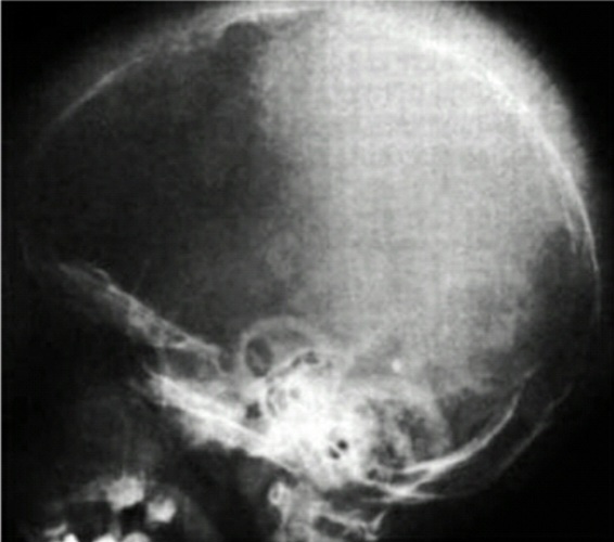
❓ Q1. Describe the radiographic findings. What is the most likely diagnosis?
✅ Model Answer:
• Hair-on-end appearance – vertical striations of thickened diploic space (classic finding).
• Thickened skull (calvarial thickening) due to marrow expansion.
• Outward bulging of skull – frontal and parietal bossing.
• Diploic space widening with radiating trabeculae.
• Diagnosis: β-thalassemia major (transfusion-dependent).
• Hair-on-end appearance – vertical striations of thickened diploic space (classic finding).
• Thickened skull (calvarial thickening) due to marrow expansion.
• Outward bulging of skull – frontal and parietal bossing.
• Diploic space widening with radiating trabeculae.
• Diagnosis: β-thalassemia major (transfusion-dependent).
❓ Q2. What is the "hair-on-end" appearance and why does it occur?
✅ Model Answer:
• Hair-on-end appearance: A classic skull X-ray finding in β-thalassemia major, characterized by vertical striations (spicules) of bone radiating from the inner table of the skull.
• Mechanism: Chronic severe anemia → ineffective erythropoiesis → massive bone marrow expansion → widening of the diploic space with compensatory thinning of the outer table → vertical trabeculae become prominent, resembling "hair standing on end".
• Also seen (less prominently) in sickle cell disease, hereditary spherocytosis, and other chronic hemolytic anemias.
• Indicates severe, longstanding marrow hyperplasia.
• Hair-on-end appearance: A classic skull X-ray finding in β-thalassemia major, characterized by vertical striations (spicules) of bone radiating from the inner table of the skull.
• Mechanism: Chronic severe anemia → ineffective erythropoiesis → massive bone marrow expansion → widening of the diploic space with compensatory thinning of the outer table → vertical trabeculae become prominent, resembling "hair standing on end".
• Also seen (less prominently) in sickle cell disease, hereditary spherocytosis, and other chronic hemolytic anemias.
• Indicates severe, longstanding marrow hyperplasia.
❓ Q3. What are the clinical features of β-thalassemia major?
✅ Model Answer:
• Pallor, fatigue, and failure to thrive – presenting in the first 6-12 months of life.
• Frontal bossing and maxillary overgrowth – "thalassemic facies" (due to marrow expansion).
• Hepatosplenomegaly – massive organomegaly (extramedullary hematopoiesis).
• Growth retardation and delayed puberty – due to chronic anemia and iron overload.
• Jaundice – from hemolysis.
• Leg ulcers – due to chronic hemolytic anemia (rare).
• Cardiac failure – due to severe anemia (if untreated) or iron overload cardiomyopathy.
• Pallor, fatigue, and failure to thrive – presenting in the first 6-12 months of life.
• Frontal bossing and maxillary overgrowth – "thalassemic facies" (due to marrow expansion).
• Hepatosplenomegaly – massive organomegaly (extramedullary hematopoiesis).
• Growth retardation and delayed puberty – due to chronic anemia and iron overload.
• Jaundice – from hemolysis.
• Leg ulcers – due to chronic hemolytic anemia (rare).
• Cardiac failure – due to severe anemia (if untreated) or iron overload cardiomyopathy.
❓ Q4. What is the pathophysiology of β-thalassemia major? Why does it cause bone changes?
✅ Model Answer:
• Pathophysiology: β-thalassemia major is caused by mutations in the β-globin gene → reduced or absent β-globin chain synthesis → imbalance of α and β chains → excess α chains precipitate in erythroid precursors → ineffective erythropoiesis and hemolysis → severe anemia.
• Bone changes mechanism: Chronic severe anemia → erythropoietin-driven marrow hyperplasia → expansion of the bone marrow cavity → thinning of cortical bone, widening of diploic space, and outward bulging of bones (frontal bossing, maxillary overgrowth).
• The skull changes (hair-on-end) are the most characteristic radiographic manifestation of this process.
• Pathophysiology: β-thalassemia major is caused by mutations in the β-globin gene → reduced or absent β-globin chain synthesis → imbalance of α and β chains → excess α chains precipitate in erythroid precursors → ineffective erythropoiesis and hemolysis → severe anemia.
• Bone changes mechanism: Chronic severe anemia → erythropoietin-driven marrow hyperplasia → expansion of the bone marrow cavity → thinning of cortical bone, widening of diploic space, and outward bulging of bones (frontal bossing, maxillary overgrowth).
• The skull changes (hair-on-end) are the most characteristic radiographic manifestation of this process.
❓ Q5. What is the differential diagnosis of the "hair-on-end" appearance on skull X-ray?
✅ Model Answer:
• β-thalassemia major – most common and classic cause.
• Sickle cell disease – less prominent hair-on-end; also shows "step-off" sign (infarction of skull bones).
• Hereditary spherocytosis – rare (if severe and untreated).
• Iron deficiency anemia – very rare (only if extremely severe and chronic).
• Congenital hemolytic anemias – other causes of chronic severe anemia.
• Primary bone disorders – Paget's disease (adults), fibrous dysplasia (different appearance).
• Key differentiating feature: Associated clinical findings (hepatosplenomegaly, anemia type) and hemoglobin electrophoresis.
• β-thalassemia major – most common and classic cause.
• Sickle cell disease – less prominent hair-on-end; also shows "step-off" sign (infarction of skull bones).
• Hereditary spherocytosis – rare (if severe and untreated).
• Iron deficiency anemia – very rare (only if extremely severe and chronic).
• Congenital hemolytic anemias – other causes of chronic severe anemia.
• Primary bone disorders – Paget's disease (adults), fibrous dysplasia (different appearance).
• Key differentiating feature: Associated clinical findings (hepatosplenomegaly, anemia type) and hemoglobin electrophoresis.
❓ Q6. What is the diagnostic workup for suspected β-thalassemia major?
✅ Model Answer:
• Complete blood count (CBC): Severe microcytic hypochromic anemia (Hb 3-7 g/dL), low MCV, low MCH.
• Peripheral smear: Target cells, nucleated red blood cells, basophilic stippling, microcytes, hypochromia.
• Hb electrophoresis: The gold standard.
- β-thalassemia major: HbF predominantly (90-100%), HbA absent (β⁰/β⁰) or reduced (β⁺/β⁺).
- HbA2: Normal or slightly elevated (2-5%).
• Serum ferritin: Elevated (due to transfusion iron overload).
• Genetic testing: For definitive diagnosis and prenatal counseling.
• X-ray of long bones and skull: Hair-on-end appearance, cortical thinning, marrow expansion.
• Complete blood count (CBC): Severe microcytic hypochromic anemia (Hb 3-7 g/dL), low MCV, low MCH.
• Peripheral smear: Target cells, nucleated red blood cells, basophilic stippling, microcytes, hypochromia.
• Hb electrophoresis: The gold standard.
- β-thalassemia major: HbF predominantly (90-100%), HbA absent (β⁰/β⁰) or reduced (β⁺/β⁺).
- HbA2: Normal or slightly elevated (2-5%).
• Serum ferritin: Elevated (due to transfusion iron overload).
• Genetic testing: For definitive diagnosis and prenatal counseling.
• X-ray of long bones and skull: Hair-on-end appearance, cortical thinning, marrow expansion.
❓ Q7. What is the management of β-thalassemia major?
✅ Model Answer:
• Chronic blood transfusions:
- Goal: Maintain Hb ≥9.5-10.5 g/dL (pre-transfusion).
- Typically every 2-4 weeks.
- Prevents severe anemia, suppresses marrow expansion, and improves growth.
• Iron chelation therapy:
- Prevents iron overload from transfusions.
- Agents: Deferoxamine (subcutaneous), Deferasirox (oral), Deferiprone (oral).
- Target ferritin <1000 ng/mL.
• Folic acid supplementation: 1 mg daily.
• Hematopoietic stem cell transplant (HSCT): Only curative option; best outcomes in young children.
• Gene therapy: Emerging treatment; promising results.
• Splenectomy: Rarely needed (if hypersplenism or massive splenomegaly).
• Chronic blood transfusions:
- Goal: Maintain Hb ≥9.5-10.5 g/dL (pre-transfusion).
- Typically every 2-4 weeks.
- Prevents severe anemia, suppresses marrow expansion, and improves growth.
• Iron chelation therapy:
- Prevents iron overload from transfusions.
- Agents: Deferoxamine (subcutaneous), Deferasirox (oral), Deferiprone (oral).
- Target ferritin <1000 ng/mL.
• Folic acid supplementation: 1 mg daily.
• Hematopoietic stem cell transplant (HSCT): Only curative option; best outcomes in young children.
• Gene therapy: Emerging treatment; promising results.
• Splenectomy: Rarely needed (if hypersplenism or massive splenomegaly).
❓ Q8. What are the complications of β-thalassemia major?
✅ Model Answer:
• Iron overload: Cardiomyopathy (most common cause of death), liver cirrhosis, endocrine dysfunction (diabetes, hypothyroidism, hypogonadism, growth retardation).
• Extramedullary hematopoiesis: Hepatosplenomegaly, paraspinal masses, pulmonary hypertension.
• Bone changes: Osteopenia, osteoporosis, pathological fractures.
• Transfusion reactions: Alloimmunization, infections (HIV, hepatitis B/C, malaria).
• Cardiac failure: Due to severe anemia (untreated) or iron overload.
• Growth retardation and delayed puberty: Due to chronic anemia and iron overload.
• Leg ulcers: Due to chronic hemolytic anemia.
• Iron overload: Cardiomyopathy (most common cause of death), liver cirrhosis, endocrine dysfunction (diabetes, hypothyroidism, hypogonadism, growth retardation).
• Extramedullary hematopoiesis: Hepatosplenomegaly, paraspinal masses, pulmonary hypertension.
• Bone changes: Osteopenia, osteoporosis, pathological fractures.
• Transfusion reactions: Alloimmunization, infections (HIV, hepatitis B/C, malaria).
• Cardiac failure: Due to severe anemia (untreated) or iron overload.
• Growth retardation and delayed puberty: Due to chronic anemia and iron overload.
• Leg ulcers: Due to chronic hemolytic anemia.
❓ Q9. How do you differentiate β-thalassemia major from β-thalassemia intermedia?
✅ Model Answer:
• β-thalassemia major:
- Transfusion-dependent.
- Hb <7 g/dL (untreated).
- Severe symptoms (failure to thrive, massive hepatosplenomegaly, marked bone changes).
- HbF >90%, HbA absent or <10%.
• β-thalassemia intermedia:
- Not transfusion-dependent (or only for complications).
- Hb 7-9 g/dL.
- Milder symptoms; may present later in childhood.
- HbF 50-90%, HbA present (10-50%).
- Less severe bone changes, less organomegaly.
• β-thalassemia major:
- Transfusion-dependent.
- Hb <7 g/dL (untreated).
- Severe symptoms (failure to thrive, massive hepatosplenomegaly, marked bone changes).
- HbF >90%, HbA absent or <10%.
• β-thalassemia intermedia:
- Not transfusion-dependent (or only for complications).
- Hb 7-9 g/dL.
- Milder symptoms; may present later in childhood.
- HbF 50-90%, HbA present (10-50%).
- Less severe bone changes, less organomegaly.
❓ Q10. A 2-year-old child with β-thalassemia major on regular transfusions develops darkening of skin, abdominal pain, and diabetes mellitus. What is the most likely cause?
✅ Model Answer:
• This is iron overload (transfusional hemosiderosis).
• Mechanism: Repeated blood transfusions lead to progressive iron accumulation in tissues (heart, liver, endocrine glands).
• Iron deposition in the pancreas causes diabetes mellitus (diabetes due to iron overload).
• Iron deposition in the skin causes hyperpigmentation.
• Liver involvement: cirrhosis, hepatomegaly.
• Management: Iron chelation therapy (deferasirox, deferoxamine, deferiprone) to reduce iron burden. Monitor ferritin and cardiac MRI (T2*).
• This is iron overload (transfusional hemosiderosis).
• Mechanism: Repeated blood transfusions lead to progressive iron accumulation in tissues (heart, liver, endocrine glands).
• Iron deposition in the pancreas causes diabetes mellitus (diabetes due to iron overload).
• Iron deposition in the skin causes hyperpigmentation.
• Liver involvement: cirrhosis, hepatomegaly.
• Management: Iron chelation therapy (deferasirox, deferoxamine, deferiprone) to reduce iron burden. Monitor ferritin and cardiac MRI (T2*).
❓ Q11. What is the role of hematopoietic stem cell transplant (HSCT) in thalassemia?
✅ Model Answer:
• HSCT is the only curative treatment for β-thalassemia major.
• Best outcomes: Young children (<5 years) with no iron overload (Pesaro class I) – survival >90%.
• Class II: Good outcomes (survival 80-85%).
• Class III: Poor outcomes (<50%) if significant iron overload (ferritin >2500, hepatomegaly, portal fibrosis).
• Donor source: HLA-matched sibling (best), matched unrelated donor, haploidentical, cord blood.
• Complications: Graft-versus-host disease (GVHD), infections, veno-occlusive disease, graft failure.
• HSCT is the only curative treatment for β-thalassemia major.
• Best outcomes: Young children (<5 years) with no iron overload (Pesaro class I) – survival >90%.
• Class II: Good outcomes (survival 80-85%).
• Class III: Poor outcomes (<50%) if significant iron overload (ferritin >2500, hepatomegaly, portal fibrosis).
• Donor source: HLA-matched sibling (best), matched unrelated donor, haploidentical, cord blood.
• Complications: Graft-versus-host disease (GVHD), infections, veno-occlusive disease, graft failure.
❓ Q12. A child with β-thalassemia major on regular transfusions develops delayed puberty and short stature. What is the most likely cause and management?
✅ Model Answer:
• This is due to endocrine dysfunction from iron overload.
• Iron deposition in the pituitary gland → growth hormone deficiency, hypogonadotrophic hypogonadism.
• Iron deposition in the thyroid → hypothyroidism.
• Management:
1. Intensify iron chelation – to reduce iron burden and prevent further endocrine damage.
2. Growth hormone therapy – if growth hormone deficiency is confirmed.
3. Thyroid hormone replacement – if hypothyroidism is present.
4. Puberty induction – sex hormone replacement for delayed puberty.
5. Regular monitoring – ferritin, endocrine function, and MRI T2* to assess iron load.
• This is due to endocrine dysfunction from iron overload.
• Iron deposition in the pituitary gland → growth hormone deficiency, hypogonadotrophic hypogonadism.
• Iron deposition in the thyroid → hypothyroidism.
• Management:
1. Intensify iron chelation – to reduce iron burden and prevent further endocrine damage.
2. Growth hormone therapy – if growth hormone deficiency is confirmed.
3. Thyroid hormone replacement – if hypothyroidism is present.
4. Puberty induction – sex hormone replacement for delayed puberty.
5. Regular monitoring – ferritin, endocrine function, and MRI T2* to assess iron load.
⚠️ Key concept: The hair‑on‑end appearance on skull X‑ray is a classic finding in β‑thalassemia major
due to marrow expansion from chronic ineffective erythropoiesis.
It represents widening of the diploic space with radiating trabeculae.
📋 Mock Test Feedback
💡 Examiner's note: Compare your answers with the model answers. In real TOACS, you would discuss these with the examiner.