FCPS Paediatrics TOACS · ECG in Pompe Disease (GSD II)
💙 4-month-old with hypotonia, macroglossia, cardiomegaly – ECG: short PR, LVH, giant QRS voltage – GAA deficiency, ERT (alglucosidase alfa)
📚 Paeds Online – paeds.online
⚕️ OBSERVED STATION · CPSP FORMAT · 8 MINUTES · SEPARATE TABS · CLINICAL SCENARIO
📖 Problem-oriented Clinical Scenario – ECG in Pompe Disease
👶🏻 Clinical Scenario (read aloud – 2 min):
A 4-month-old infant is referred to the pediatric cardiology clinic because of cardiomegaly detected on a chest X-ray obtained during an evaluation for failure to thrive. The mother reports that the infant has been floppy since birth, has poor feeding, and has difficulty breathing during feeds. She also notes that the infant's tongue appears large and protrudes. There is no family history of heart disease or metabolic disorders.
Examination: Vital signs: HR 110 bpm, BP 95/60 mm Hg, RR 45/min, SpO2 95% on room air. The infant is hypotonic with poor head control. Examination reveals macroglossia, a prominent left ventricular heave, and a soft ejection murmur at the left lower sternal border. There is no hepatomegaly or peripheral edema. Chest X-ray shows massive cardiomegaly with a globular heart shape.
ECG (obtained in clinic):
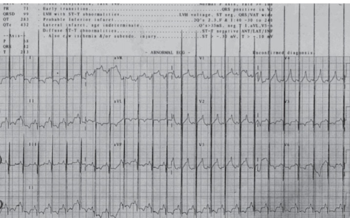 Task for the candidate: You are the pediatric cardiologist. Evaluate this infant, interpret the ECG findings (short PR interval, giant QRS voltages, LVH), discuss the differential diagnosis of the clinical features (Pompe disease – GSD II), formulate a diagnostic plan (GAA enzyme assay, genetic testing), and provide counseling to the parents about the diagnosis, treatment (enzyme replacement therapy – alglucosidase alfa), and prognosis.
A 4-month-old infant is referred to the pediatric cardiology clinic because of cardiomegaly detected on a chest X-ray obtained during an evaluation for failure to thrive. The mother reports that the infant has been floppy since birth, has poor feeding, and has difficulty breathing during feeds. She also notes that the infant's tongue appears large and protrudes. There is no family history of heart disease or metabolic disorders.
Examination: Vital signs: HR 110 bpm, BP 95/60 mm Hg, RR 45/min, SpO2 95% on room air. The infant is hypotonic with poor head control. Examination reveals macroglossia, a prominent left ventricular heave, and a soft ejection murmur at the left lower sternal border. There is no hepatomegaly or peripheral edema. Chest X-ray shows massive cardiomegaly with a globular heart shape.
ECG (obtained in clinic):
Figure: ECG shows short PR interval (0.07 sec), giant QRS voltages in V5-V6 (LVH), deep narrow Q waves in lateral leads, and ST-T wave changes.
💡 Examiner instruction (interactive): This is a case of Pompe Disease (Glycogen Storage Disease Type II) – an autosomal recessive disorder caused by deficiency of acid α-glucosidase (GAA). The candidate must recognize the classic ECG findings: short PR interval (often <0.08 seconds), giant QRS voltages (left ventricular hypertrophy), deep Q waves in leads I, aVL, V5-V6 (pseudo-infarct pattern), and ST-T changes. The candidate should also recognize the clinical features (infantile-onset: hypotonia, macroglossia, cardiomegaly, hypertrophic cardiomyopathy, failure to thrive) and understand that enzyme replacement therapy (alglucosidase alfa) is the standard of care. The candidate should discuss the diagnostic workup (GAA enzyme activity in dried blood spot or muscle biopsy, GAA gene sequencing) and the importance of early treatment to prevent progression of cardiomyopathy and respiratory failure.
🔍 Examiner Questions (interactive) – Click to reveal model answers
❓ Q1 (Examiner): “Describe the ECG findings in this infant. What are the characteristic ECG features of Pompe disease?”
✅ Candidate's answer:
• Characteristic ECG findings in Pompe disease (infantile-onset):
1️⃣ Short PR interval – less than 0.08 seconds in infants (normal 0.08-0.12 sec). This is a hallmark finding.
2️⃣ Giant QRS voltages – severe left ventricular hypertrophy (LVH) with tall R waves in V5-V6 (often >98th percentile).
3️⃣ Deep narrow Q waves – in leads I, aVL, V5-V6 (pseudo-infarct pattern).
4️⃣ ST segment depression and T-wave inversion in lateral leads (strain pattern).
5️⃣ Left atrial enlargement – broad, notched P waves (P mitrale) in lead II.
6️⃣ Left axis deviation – may be present.
• Why these changes occur:
- Glycogen accumulation in the myocardium leads to massive ventricular hypertrophy with a characteristic "electric shock" appearance on ECG.
- The short PR interval is due to accelerated AV conduction (often associated with glycogen storage diseases, especially Pompe).
- The deep Q waves are due to septal hypertrophy (pseudo-infarct pattern).
• Differential diagnosis: Other causes of short PR + LVH: Fabry disease, Danon disease, PRKAG2 syndrome, severe anemia, and thyrotoxicosis.
• Characteristic ECG findings in Pompe disease (infantile-onset):
1️⃣ Short PR interval – less than 0.08 seconds in infants (normal 0.08-0.12 sec). This is a hallmark finding.
2️⃣ Giant QRS voltages – severe left ventricular hypertrophy (LVH) with tall R waves in V5-V6 (often >98th percentile).
3️⃣ Deep narrow Q waves – in leads I, aVL, V5-V6 (pseudo-infarct pattern).
4️⃣ ST segment depression and T-wave inversion in lateral leads (strain pattern).
5️⃣ Left atrial enlargement – broad, notched P waves (P mitrale) in lead II.
6️⃣ Left axis deviation – may be present.
• Why these changes occur:
- Glycogen accumulation in the myocardium leads to massive ventricular hypertrophy with a characteristic "electric shock" appearance on ECG.
- The short PR interval is due to accelerated AV conduction (often associated with glycogen storage diseases, especially Pompe).
- The deep Q waves are due to septal hypertrophy (pseudo-infarct pattern).
• Differential diagnosis: Other causes of short PR + LVH: Fabry disease, Danon disease, PRKAG2 syndrome, severe anemia, and thyrotoxicosis.
❓ Q2 (Examiner): “What are the clinical features of infantile-onset Pompe disease? Why does this infant have macroglossia and hypotonia?”
✅ Candidate's answer:
• Clinical features of infantile-onset Pompe disease (GSD II):
- Onset: Typically within the first 2-6 months of life.
- Cardiac: Massive hypertrophic cardiomyopathy (cardiomegaly, heart failure).
- Neuromuscular: Severe hypotonia (floppy infant), poor head control, weak cry, difficulty feeding.
- Oropharyngeal: Macroglossia (enlarged tongue due to glycogen accumulation).
- Respiratory: Respiratory insufficiency, recurrent infections, respiratory failure.
- Musculoskeletal: Delayed motor milestones, progressive weakness.
- Hepatomegaly: Mild to moderate (less prominent than in GSD I).
- Dysphagia – due to muscle weakness and macroglossia.
• Why macroglossia and hypotonia?
- Macroglossia: Glycogen accumulation in the tongue muscle leads to enlargement.
- Hypotonia: Glycogen accumulation in skeletal muscle (especially type II fibers) causes muscle weakness and hypotonia.
- Pathophysiology: Deficiency of acid α-glucosidase (GAA) → failure to degrade glycogen to glucose → glycogen accumulation in lysosomes → cellular dysfunction in cardiac, skeletal, and smooth muscle.
• Clinical features of infantile-onset Pompe disease (GSD II):
- Onset: Typically within the first 2-6 months of life.
- Cardiac: Massive hypertrophic cardiomyopathy (cardiomegaly, heart failure).
- Neuromuscular: Severe hypotonia (floppy infant), poor head control, weak cry, difficulty feeding.
- Oropharyngeal: Macroglossia (enlarged tongue due to glycogen accumulation).
- Respiratory: Respiratory insufficiency, recurrent infections, respiratory failure.
- Musculoskeletal: Delayed motor milestones, progressive weakness.
- Hepatomegaly: Mild to moderate (less prominent than in GSD I).
- Dysphagia – due to muscle weakness and macroglossia.
• Why macroglossia and hypotonia?
- Macroglossia: Glycogen accumulation in the tongue muscle leads to enlargement.
- Hypotonia: Glycogen accumulation in skeletal muscle (especially type II fibers) causes muscle weakness and hypotonia.
- Pathophysiology: Deficiency of acid α-glucosidase (GAA) → failure to degrade glycogen to glucose → glycogen accumulation in lysosomes → cellular dysfunction in cardiac, skeletal, and smooth muscle.
❓ Q3 (Examiner): “How is Pompe disease diagnosed? What is the role of GAA enzyme assay and genetic testing?”
✅ Candidate's answer:
• Diagnostic approach for Pompe disease:
1️⃣ GAA enzyme activity assay: The gold standard. Can be performed on:
- Dried blood spot (DBS): First-line screening test.
- Leukocytes, fibroblasts, or muscle biopsy.
- Reduced GAA activity (<10% of normal) confirms the diagnosis of Pompe disease.
2️⃣ Genetic testing: Sequencing of the GAA gene (chromosome 17q25) – identifies the causative mutations. This is important for confirming the diagnosis, genetic counseling, and prenatal diagnosis.
3️⃣ Newborn screening: Dried blood spot GAA assay is increasingly used in newborn screening programs.
4️⃣ Other tests:
- Echocardiogram: Shows severe concentric LVH, with normal or hyperdynamic systolic function in early stages.
- CK: May be elevated (due to muscle involvement).
- ECG: Short PR interval, LVH, giant QRS voltages.
- Muscle biopsy: Glycogen accumulation (PAS-positive vacuoles) in muscle tissue (less commonly used now).
• Diagnostic approach for Pompe disease:
1️⃣ GAA enzyme activity assay: The gold standard. Can be performed on:
- Dried blood spot (DBS): First-line screening test.
- Leukocytes, fibroblasts, or muscle biopsy.
- Reduced GAA activity (<10% of normal) confirms the diagnosis of Pompe disease.
2️⃣ Genetic testing: Sequencing of the GAA gene (chromosome 17q25) – identifies the causative mutations. This is important for confirming the diagnosis, genetic counseling, and prenatal diagnosis.
3️⃣ Newborn screening: Dried blood spot GAA assay is increasingly used in newborn screening programs.
4️⃣ Other tests:
- Echocardiogram: Shows severe concentric LVH, with normal or hyperdynamic systolic function in early stages.
- CK: May be elevated (due to muscle involvement).
- ECG: Short PR interval, LVH, giant QRS voltages.
- Muscle biopsy: Glycogen accumulation (PAS-positive vacuoles) in muscle tissue (less commonly used now).
❓ Q4 (Examiner): “What is the treatment for Pompe disease? What is the role of enzyme replacement therapy (ERT)?”
✅ Candidate's answer:
• Treatment of Pompe disease:
1️⃣ Enzyme replacement therapy (ERT):
- Alglucosidase alfa (Myozyme): Recombinant human acid α-glucosidase.
- Dose: 20 mg/kg IV every 2 weeks.
- Infusion: Given via IV infusion over 2-4 hours. Life-long therapy.
- Effectiveness: Improves cardiac function, reduces LVH, improves survival and motor outcomes, especially when started early (before 6 months of age).
2️⃣ Supportive care:
- Respiratory support: Non-invasive ventilation (BiPAP), mechanical ventilation if needed.
- Nutritional support: High-calorie diet, feeding tube (G-tube) if dysphagia.
- Physical therapy: To maintain muscle function.
3️⃣ Clinical trials: Gene therapy is under investigation.
4️⃣ Prognosis: Without treatment, infantile Pompe disease is fatal by 1 year (due to cardiorespiratory failure). With ERT, survival and quality of life are significantly improved, but patients may develop progressive muscle weakness (anti-GAA antibodies can reduce efficacy).
• Treatment of Pompe disease:
1️⃣ Enzyme replacement therapy (ERT):
- Alglucosidase alfa (Myozyme): Recombinant human acid α-glucosidase.
- Dose: 20 mg/kg IV every 2 weeks.
- Infusion: Given via IV infusion over 2-4 hours. Life-long therapy.
- Effectiveness: Improves cardiac function, reduces LVH, improves survival and motor outcomes, especially when started early (before 6 months of age).
2️⃣ Supportive care:
- Respiratory support: Non-invasive ventilation (BiPAP), mechanical ventilation if needed.
- Nutritional support: High-calorie diet, feeding tube (G-tube) if dysphagia.
- Physical therapy: To maintain muscle function.
3️⃣ Clinical trials: Gene therapy is under investigation.
4️⃣ Prognosis: Without treatment, infantile Pompe disease is fatal by 1 year (due to cardiorespiratory failure). With ERT, survival and quality of life are significantly improved, but patients may develop progressive muscle weakness (anti-GAA antibodies can reduce efficacy).
❓ Q5 (Examiner): “What is the differential diagnosis of infantile hypotonia, macroglossia, and cardiomyopathy?”
✅ Candidate's answer:
• Differential diagnosis of infantile hypotonia, macroglossia, and cardiomyopathy:
1️⃣ Pompe disease (GSD II) – most classic association.
2️⃣ Danon disease (LAMP2 deficiency) – X-linked, HCM + WPW + mental retardation + skeletal myopathy.
3️⃣ PRKAG2 syndrome – HCM + WPW + conduction disease (glycogen storage).
4️⃣ Fabry disease (X-linked) – HCM + angiokeratomas + renal failure + stroke.
5️⃣ Mitochondrial disorders – can cause cardiomyopathy and hypotonia.
6️⃣ Congenital hypothyroidism – macroglossia, hypotonia, but no cardiomyopathy.
7️⃣ Beckwith-Wiedemann syndrome – macroglossia, visceromegaly, but no cardiomyopathy.
8️⃣ Noonan syndrome – HCM, but no macroglossia typically.
9️⃣ Congenital myopathies – hypotonia, but no cardiomyopathy.
• Key differentiating features: ECG (short PR + giant QRS = Pompe), associated WPW (Danon, PRKAG2), pattern of inheritance (X-linked for Danon/Fabry, AR for Pompe).
• Differential diagnosis of infantile hypotonia, macroglossia, and cardiomyopathy:
1️⃣ Pompe disease (GSD II) – most classic association.
2️⃣ Danon disease (LAMP2 deficiency) – X-linked, HCM + WPW + mental retardation + skeletal myopathy.
3️⃣ PRKAG2 syndrome – HCM + WPW + conduction disease (glycogen storage).
4️⃣ Fabry disease (X-linked) – HCM + angiokeratomas + renal failure + stroke.
5️⃣ Mitochondrial disorders – can cause cardiomyopathy and hypotonia.
6️⃣ Congenital hypothyroidism – macroglossia, hypotonia, but no cardiomyopathy.
7️⃣ Beckwith-Wiedemann syndrome – macroglossia, visceromegaly, but no cardiomyopathy.
8️⃣ Noonan syndrome – HCM, but no macroglossia typically.
9️⃣ Congenital myopathies – hypotonia, but no cardiomyopathy.
• Key differentiating features: ECG (short PR + giant QRS = Pompe), associated WPW (Danon, PRKAG2), pattern of inheritance (X-linked for Danon/Fabry, AR for Pompe).
❓ Q6 (Examiner): “Why is the PR interval short in Pompe disease? What other conditions cause a short PR interval?”
✅ Candidate's answer:
• Mechanism of short PR in Pompe: Glycogen accumulation in the myocardium and conduction system leads to accelerated atrioventricular (AV) conduction, resulting in a short PR interval. This is a characteristic ECG feature.
• Other causes of short PR interval (<0.08 sec in infants):
1️⃣ Wolff-Parkinson-White (WPW) syndrome – short PR + delta wave + wide QRS.
2️⃣ Lown-Ganong-Levine (LGL) syndrome – short PR without delta wave (James fibers).
3️⃣ Glycogen storage diseases: Pompe (GSD II), Danon disease (LAMP2), PRKAG2 syndrome.
4️⃣ Thyrotoxicosis – increases AV conduction.
5️⃣ Hyperkalemia – can shorten PR (but usually with peaked T waves).
6️⃣ Severe anemia – increased stroke volume → accelerated conduction.
7️⃣ Digitalis toxicity.
• Differentiation: In Pompe, there is no delta wave, and the QRS is widened due to LVH (giant QRS voltages).
• Mechanism of short PR in Pompe: Glycogen accumulation in the myocardium and conduction system leads to accelerated atrioventricular (AV) conduction, resulting in a short PR interval. This is a characteristic ECG feature.
• Other causes of short PR interval (<0.08 sec in infants):
1️⃣ Wolff-Parkinson-White (WPW) syndrome – short PR + delta wave + wide QRS.
2️⃣ Lown-Ganong-Levine (LGL) syndrome – short PR without delta wave (James fibers).
3️⃣ Glycogen storage diseases: Pompe (GSD II), Danon disease (LAMP2), PRKAG2 syndrome.
4️⃣ Thyrotoxicosis – increases AV conduction.
5️⃣ Hyperkalemia – can shorten PR (but usually with peaked T waves).
6️⃣ Severe anemia – increased stroke volume → accelerated conduction.
7️⃣ Digitalis toxicity.
• Differentiation: In Pompe, there is no delta wave, and the QRS is widened due to LVH (giant QRS voltages).
❓ Q7 (Examiner): “What causes the giant QRS voltages and deep Q waves in Pompe disease? What is the differential diagnosis of giant QRS voltages?”
✅ Candidate's answer:
• Giant QRS voltages: Due to massive left ventricular hypertrophy from glycogen accumulation in the myocardium. The thickness of the ventricular wall increases the electrical forces, producing tall R waves in the left precordial leads (V5-V6) and deep S waves in V1-V2.
• Deep Q waves: Due to septal hypertrophy – the hypertrophied septum produces a pseudo-infarct pattern (deep narrow Q waves in lateral leads).
• Differential diagnosis of giant QRS voltages (LVH):
1️⃣ Hypertrophic cardiomyopathy (HCM): Sarcomeric (MYH7, MYBPC3).
2️⃣ Pompe disease (GSD II).
3️⃣ Danon disease (LAMP2).
4️⃣ PRKAG2 syndrome.
5️⃣ Noonan syndrome (RASopathy).
6️⃣ Friedreich ataxia.
7️⃣ Aortic stenosis.
8️⃣ Coarctation of aorta.
• Key to differentiation: In Pompe, the short PR + giant QRS + deep Q waves + infantile presentation (hypotonia, macroglossia) is highly characteristic.
• Giant QRS voltages: Due to massive left ventricular hypertrophy from glycogen accumulation in the myocardium. The thickness of the ventricular wall increases the electrical forces, producing tall R waves in the left precordial leads (V5-V6) and deep S waves in V1-V2.
• Deep Q waves: Due to septal hypertrophy – the hypertrophied septum produces a pseudo-infarct pattern (deep narrow Q waves in lateral leads).
• Differential diagnosis of giant QRS voltages (LVH):
1️⃣ Hypertrophic cardiomyopathy (HCM): Sarcomeric (MYH7, MYBPC3).
2️⃣ Pompe disease (GSD II).
3️⃣ Danon disease (LAMP2).
4️⃣ PRKAG2 syndrome.
5️⃣ Noonan syndrome (RASopathy).
6️⃣ Friedreich ataxia.
7️⃣ Aortic stenosis.
8️⃣ Coarctation of aorta.
• Key to differentiation: In Pompe, the short PR + giant QRS + deep Q waves + infantile presentation (hypotonia, macroglossia) is highly characteristic.
❓ Q8 (Examiner): “What is the natural history of untreated infantile Pompe disease? How does ERT change the prognosis?”
✅ Candidate's answer:
• Untreated infantile Pompe disease:
- Natural history: Rapidly progressive hypertrophic cardiomyopathy, severe hypotonia, macroglossia, and respiratory insufficiency.
- Death: Usually by 1 year of age (median survival ~8-12 months) due to cardiorespiratory failure.
- Progressive weakness: Leads to failure to thrive, feeding difficulties, and recurrent respiratory infections.
• With enzyme replacement therapy (ERT):
- Cardiac outcomes: LVH reverses or stabilizes, heart failure improves.
- Survival: Significant improvement – many patients survive into childhood and beyond.
- Motor outcomes: Delayed but improved compared to untreated; patients may achieve sitting, standing, and walking (with variable degrees).
- Challenges: Progressive muscle weakness may continue (especially if anti-GAA antibodies develop), requiring respiratory support and feeding tubes.
- Optimal timing: Early initiation of ERT (before 6 months of age, ideally before symptoms) is associated with the best outcomes.
• Untreated infantile Pompe disease:
- Natural history: Rapidly progressive hypertrophic cardiomyopathy, severe hypotonia, macroglossia, and respiratory insufficiency.
- Death: Usually by 1 year of age (median survival ~8-12 months) due to cardiorespiratory failure.
- Progressive weakness: Leads to failure to thrive, feeding difficulties, and recurrent respiratory infections.
• With enzyme replacement therapy (ERT):
- Cardiac outcomes: LVH reverses or stabilizes, heart failure improves.
- Survival: Significant improvement – many patients survive into childhood and beyond.
- Motor outcomes: Delayed but improved compared to untreated; patients may achieve sitting, standing, and walking (with variable degrees).
- Challenges: Progressive muscle weakness may continue (especially if anti-GAA antibodies develop), requiring respiratory support and feeding tubes.
- Optimal timing: Early initiation of ERT (before 6 months of age, ideally before symptoms) is associated with the best outcomes.
❓ Q9 (Examiner): “What are the echocardiographic findings in Pompe disease? How does it differ from other causes of HCM?”
✅ Candidate's answer:
• Echocardiographic findings in Pompe disease:
- Severe concentric left ventricular hypertrophy (LVH): LV wall thickness is markedly increased (z-score >5).
- Normal or hyperdynamic LV systolic function in early stages (EF >60%).
- Diastolic dysfunction – impaired relaxation (E/A reversal).
- Left ventricular outflow tract (LVOT) obstruction: May be present (but less common than in sarcomeric HCM).
- Biventricular hypertrophy: RV involvement is common.
- Valvular abnormalities: Mitral valve thickening may occur (due to glycogen deposition).
- Progression: LV function may deteriorate over time if untreated.
• Differences from sarcomeric HCM:
- Pompe: More severe concentric hypertrophy, early onset (infancy), associated with hypotonia and macroglossia.
- Sarcomeric HCM: Asymmetric septal hypertrophy, LVOT obstruction more common, typically later onset (childhood/adolescence).
• Echocardiographic findings in Pompe disease:
- Severe concentric left ventricular hypertrophy (LVH): LV wall thickness is markedly increased (z-score >5).
- Normal or hyperdynamic LV systolic function in early stages (EF >60%).
- Diastolic dysfunction – impaired relaxation (E/A reversal).
- Left ventricular outflow tract (LVOT) obstruction: May be present (but less common than in sarcomeric HCM).
- Biventricular hypertrophy: RV involvement is common.
- Valvular abnormalities: Mitral valve thickening may occur (due to glycogen deposition).
- Progression: LV function may deteriorate over time if untreated.
• Differences from sarcomeric HCM:
- Pompe: More severe concentric hypertrophy, early onset (infancy), associated with hypotonia and macroglossia.
- Sarcomeric HCM: Asymmetric septal hypertrophy, LVOT obstruction more common, typically later onset (childhood/adolescence).
❓ Q10 (Examiner): “What is the role of newborn screening for Pompe disease? Why is early detection important?”
✅ Candidate's answer:
• Newborn screening for Pompe disease: Many states/countries have implemented newborn screening for Pompe disease using dried blood spot (DBS) GAA enzyme assay.
• Why early detection is important:
- Early initiation of ERT: Starting alglucosidase alfa before 6 months of age (ideally in the first few weeks of life) significantly improves outcomes.
- Prevention of irreversible cardiomyopathy: ERT can reverse or halt the progression of LVH.
- Improved motor outcomes: Early treatment leads to better motor milestones (sitting, walking).
- Reduced mortality: Without newborn screening, many infants are diagnosed late (after symptoms develop), leading to poor outcomes.
- Cost-effectiveness: Early detection reduces the need for intensive care and improves quality of life.
• Newborn screening for Pompe disease: Many states/countries have implemented newborn screening for Pompe disease using dried blood spot (DBS) GAA enzyme assay.
• Why early detection is important:
- Early initiation of ERT: Starting alglucosidase alfa before 6 months of age (ideally in the first few weeks of life) significantly improves outcomes.
- Prevention of irreversible cardiomyopathy: ERT can reverse or halt the progression of LVH.
- Improved motor outcomes: Early treatment leads to better motor milestones (sitting, walking).
- Reduced mortality: Without newborn screening, many infants are diagnosed late (after symptoms develop), leading to poor outcomes.
- Cost-effectiveness: Early detection reduces the need for intensive care and improves quality of life.
❓ Q11 (Examiner): “What is the inheritance pattern of Pompe disease? What is the recurrence risk for future pregnancies?”
✅ Candidate's answer:
• Inheritance pattern: Autosomal recessive (parents are carriers, usually asymptomatic).
• Gene: GAA gene located on chromosome 17q25.
• Recurrence risk:
- For each future pregnancy, there is a 25% chance of having an affected child (inheriting both mutant alleles).
- 50% chance of the child being a carrier (heterozygote).
- 25% chance of being unaffected (homozygous normal).
• Prenatal diagnosis: Available through chorionic villus sampling (CVS) or amniocentesis to test for GAA mutations or enzyme activity.
• Genetic counseling: Offer genetic testing to the parents (carrier testing) and to other family members. Prenatal diagnosis should be discussed. Preimplantation genetic diagnosis (PGD) is also an option.
• Inheritance pattern: Autosomal recessive (parents are carriers, usually asymptomatic).
• Gene: GAA gene located on chromosome 17q25.
• Recurrence risk:
- For each future pregnancy, there is a 25% chance of having an affected child (inheriting both mutant alleles).
- 50% chance of the child being a carrier (heterozygote).
- 25% chance of being unaffected (homozygous normal).
• Prenatal diagnosis: Available through chorionic villus sampling (CVS) or amniocentesis to test for GAA mutations or enzyme activity.
• Genetic counseling: Offer genetic testing to the parents (carrier testing) and to other family members. Prenatal diagnosis should be discussed. Preimplantation genetic diagnosis (PGD) is also an option.
❓ Q12 (Examiner): “What are the complications of enzyme replacement therapy in Pompe disease? How are they managed?”
✅ Candidate's answer:
• Complications of ERT (alglucosidase alfa):
1️⃣ Infusion-related reactions: Fever, rash, urticaria, flushing, and anaphylaxis. Premedication with antihistamines and corticosteroids is often given.
2️⃣ Anti-GAA antibody formation: Some patients develop antibodies that may reduce the efficacy of ERT. Immunomodulation (e.g., with rituximab, methotrexate) may be considered in some centers.
3️⃣ Progressive muscle weakness: Despite ERT, some patients develop respiratory muscle weakness and require ventilatory support. Physical therapy and respiratory support are essential.
4️⃣ Cardiac complications: While LVH improves, arrhythmias may persist.
5️⃣ Hepatotoxicity: Liver enzymes should be monitored (rare).
• Management:
- Premedication for infusions.
- Monitor for antibodies and adjust therapy.
- Multidisciplinary care (cardiologist, pulmonologist, neurologist, geneticist).
• Complications of ERT (alglucosidase alfa):
1️⃣ Infusion-related reactions: Fever, rash, urticaria, flushing, and anaphylaxis. Premedication with antihistamines and corticosteroids is often given.
2️⃣ Anti-GAA antibody formation: Some patients develop antibodies that may reduce the efficacy of ERT. Immunomodulation (e.g., with rituximab, methotrexate) may be considered in some centers.
3️⃣ Progressive muscle weakness: Despite ERT, some patients develop respiratory muscle weakness and require ventilatory support. Physical therapy and respiratory support are essential.
4️⃣ Cardiac complications: While LVH improves, arrhythmias may persist.
5️⃣ Hepatotoxicity: Liver enzymes should be monitored (rare).
• Management:
- Premedication for infusions.
- Monitor for antibodies and adjust therapy.
- Multidisciplinary care (cardiologist, pulmonologist, neurologist, geneticist).
❓ Q13 (Examiner): “What other glycogen storage diseases can cause cardiomyopathy? How do they differ from Pompe?”
✅ Candidate's answer:
• Other GSDs with cardiomyopathy:
1️⃣ Danon disease (LAMP2): X-linked, HCM + WPW + mental retardation + skeletal myopathy. ECG: short PR, delta wave (WPW), LVH.
2️⃣ PRKAG2 syndrome: Autosomal dominant, HCM + WPW + conduction disease. ECG: short PR, delta wave, LVH, progressive AV block.
3️⃣ GSD III (Cori/Forbes, debrancher deficiency): Can cause cardiomyopathy (hypertrophic or dilated) in addition to hepatomegaly and hypoglycemia.
4️⃣ GSD IV (Andersen, branching enzyme deficiency): Liver cirrhosis, but cardiomyopathy can occur.
5️⃣ Pompe (GSD II): Most severe cardiomyopathy in infancy, with characteristic ECG (short PR, giant QRS, no delta wave).
• Key differences:
- Danon/PRKAG2: Associated with WPW (delta wave), mental retardation (Danon).
- Pompe: No delta wave, infantile presentation with macroglossia and hypotonia.
- GSD III: Hepatomegaly, hypoglycemia, muscle weakness, cardiomyopathy (later onset).
• Other GSDs with cardiomyopathy:
1️⃣ Danon disease (LAMP2): X-linked, HCM + WPW + mental retardation + skeletal myopathy. ECG: short PR, delta wave (WPW), LVH.
2️⃣ PRKAG2 syndrome: Autosomal dominant, HCM + WPW + conduction disease. ECG: short PR, delta wave, LVH, progressive AV block.
3️⃣ GSD III (Cori/Forbes, debrancher deficiency): Can cause cardiomyopathy (hypertrophic or dilated) in addition to hepatomegaly and hypoglycemia.
4️⃣ GSD IV (Andersen, branching enzyme deficiency): Liver cirrhosis, but cardiomyopathy can occur.
5️⃣ Pompe (GSD II): Most severe cardiomyopathy in infancy, with characteristic ECG (short PR, giant QRS, no delta wave).
• Key differences:
- Danon/PRKAG2: Associated with WPW (delta wave), mental retardation (Danon).
- Pompe: No delta wave, infantile presentation with macroglossia and hypotonia.
- GSD III: Hepatomegaly, hypoglycemia, muscle weakness, cardiomyopathy (later onset).
❓ Q14 (Examiner): “The parents are very anxious about their infant's diagnosis. How will you counsel them?”
✅ Candidate's structured answer:
• “Your baby has a condition called Pompe disease – a rare genetic disorder where the body cannot break down a type of sugar (glycogen) in the muscle cells, including the heart muscle.”
• “This causes the heart to become very thick (hypertrophic cardiomyopathy) and the muscles to be weak (hypotonia). The ECG shows characteristic changes – a very short PR interval and giant QRS voltages – which are classic for this condition.”
• “The good news is that we have a very effective treatment called enzyme replacement therapy (ERT) – a medication given through an IV every 2 weeks. It replaces the missing enzyme and can significantly improve heart function, muscle strength, and survival.”
• “The earlier we start treatment, the better the outcomes. Your baby will need lifelong ERT, and we will also provide supportive care – respiratory support, nutrition, and physical therapy.”
• “We will monitor the heart with regular echocardiograms and ECGs. Most children with Pompe disease who are treated early can live into adulthood.”
• “This is an autosomal recessive condition, meaning both you and your partner are carriers. For future pregnancies, there is a 25% recurrence risk. We can discuss prenatal testing options.”
• “You are not alone – we have a team of specialists who will support you and your baby every step of the way.”
• “Your baby has a condition called Pompe disease – a rare genetic disorder where the body cannot break down a type of sugar (glycogen) in the muscle cells, including the heart muscle.”
• “This causes the heart to become very thick (hypertrophic cardiomyopathy) and the muscles to be weak (hypotonia). The ECG shows characteristic changes – a very short PR interval and giant QRS voltages – which are classic for this condition.”
• “The good news is that we have a very effective treatment called enzyme replacement therapy (ERT) – a medication given through an IV every 2 weeks. It replaces the missing enzyme and can significantly improve heart function, muscle strength, and survival.”
• “The earlier we start treatment, the better the outcomes. Your baby will need lifelong ERT, and we will also provide supportive care – respiratory support, nutrition, and physical therapy.”
• “We will monitor the heart with regular echocardiograms and ECGs. Most children with Pompe disease who are treated early can live into adulthood.”
• “This is an autosomal recessive condition, meaning both you and your partner are carriers. For future pregnancies, there is a 25% recurrence risk. We can discuss prenatal testing options.”
• “You are not alone – we have a team of specialists who will support you and your baby every step of the way.”
🗣️ Examiner's probing / high-yield points (ECG in Pompe Disease):
• "What are the classic ECG findings in Pompe disease?" → Short PR, giant QRS voltages, LVH, deep Q waves.
• "What is the enzyme deficiency in Pompe disease?" → Acid α-glucosidase (GAA).
• "What is the treatment for Pompe disease?" → Enzyme replacement therapy (alglucosidase alfa).
• "What is the inheritance pattern?" → Autosomal recessive.
• "What is the recurrence risk?" → 25% for each pregnancy.
• "What is the differential diagnosis for short PR + LVH?" → Danon disease, PRKAG2, Fabry, HCM.
• "What is the natural history without treatment?" → Death by 1 year (cardiorespiratory failure).
• "Why is newborn screening important?" → Early ERT improves outcomes.
• "What is the mechanism of macroglossia?" → Glycogen accumulation in tongue.
• "What is the mechanism of hypotonia?" → Glycogen accumulation in skeletal muscle.
• "What are the classic ECG findings in Pompe disease?" → Short PR, giant QRS voltages, LVH, deep Q waves.
• "What is the enzyme deficiency in Pompe disease?" → Acid α-glucosidase (GAA).
• "What is the treatment for Pompe disease?" → Enzyme replacement therapy (alglucosidase alfa).
• "What is the inheritance pattern?" → Autosomal recessive.
• "What is the recurrence risk?" → 25% for each pregnancy.
• "What is the differential diagnosis for short PR + LVH?" → Danon disease, PRKAG2, Fabry, HCM.
• "What is the natural history without treatment?" → Death by 1 year (cardiorespiratory failure).
• "Why is newborn screening important?" → Early ERT improves outcomes.
• "What is the mechanism of macroglossia?" → Glycogen accumulation in tongue.
• "What is the mechanism of hypotonia?" → Glycogen accumulation in skeletal muscle.
📘 ECG in Pompe Disease – Core Revision for TOACS
💙 Definition
Pompe disease (GSD II) – autosomal recessive deficiency of acid α-glucosidase (GAA). Glycogen accumulation in lysosomes → cardiomyopathy, hypotonia, macroglossia.
Pompe disease (GSD II) – autosomal recessive deficiency of acid α-glucosidase (GAA). Glycogen accumulation in lysosomes → cardiomyopathy, hypotonia, macroglossia.
📊 Classic ECG Findings
Short PR interval (<0.08 sec), giant QRS voltages (LVH), deep narrow Q waves (lateral leads), ST-T changes, left atrial enlargement.
Short PR interval (<0.08 sec), giant QRS voltages (LVH), deep narrow Q waves (lateral leads), ST-T changes, left atrial enlargement.
🔍 Clinical Features
Infantile-onset: massive HCM, hypotonia, macroglossia, failure to thrive, respiratory failure. Without treatment, death by 1 year.
Infantile-onset: massive HCM, hypotonia, macroglossia, failure to thrive, respiratory failure. Without treatment, death by 1 year.
🩺 Diagnosis
GAA enzyme assay (dried blood spot – newborn screening). Genetic testing (GAA gene). Echocardiogram: severe concentric LVH.
GAA enzyme assay (dried blood spot – newborn screening). Genetic testing (GAA gene). Echocardiogram: severe concentric LVH.
💊 Treatment
Enzyme replacement therapy (alglucosidase alfa) 20 mg/kg IV every 2 weeks. Early initiation (<6 months) improves outcomes.
Enzyme replacement therapy (alglucosidase alfa) 20 mg/kg IV every 2 weeks. Early initiation (<6 months) improves outcomes.
🧬 Genetics
Autosomal recessive. GAA gene (17q25). Recurrence risk: 25%. Prenatal diagnosis available.
Autosomal recessive. GAA gene (17q25). Recurrence risk: 25%. Prenatal diagnosis available.
⭐ High-yield pearls for TOACS (ECG in Pompe Disease):
• ECG: Short PR + giant QRS + LVH + deep Q waves.
• Pompe = GSD II = acid α-glucosidase deficiency.
• Clinical triad: Cardiomyopathy + hypotonia + macroglossia.
• Treatment: ERT (alglucosidase alfa).
• Inheritance: Autosomal recessive (25% recurrence risk).
• Untreated: death by 1 year.
• Differential: Danon (WPW + mental retardation), PRKAG2 (WPW + conduction disease).
• ECG: Short PR + giant QRS + LVH + deep Q waves.
• Pompe = GSD II = acid α-glucosidase deficiency.
• Clinical triad: Cardiomyopathy + hypotonia + macroglossia.
• Treatment: ERT (alglucosidase alfa).
• Inheritance: Autosomal recessive (25% recurrence risk).
• Untreated: death by 1 year.
• Differential: Danon (WPW + mental retardation), PRKAG2 (WPW + conduction disease).
🗣️ Candidate's role-play & examiner feedback
💬 To the candidate (role‑play): You will be asked the 14 questions from the Examiner Q&A tab. This station tests knowledge of ECG in Pompe Disease – classic findings (short PR, giant QRS, LVH, deep Q waves), clinical features (hypotonia, macroglossia, cardiomyopathy), diagnosis (GAA enzyme assay), treatment (ERT), and genetics (autosomal recessive). Provide empathetic counseling to parents about the diagnosis, treatment, and prognosis.
📝 Examiner Marking Grid (ECG in Pompe Disease – TOACS station):
- ✅ Interprets ECG: short PR, giant QRS, LVH, deep Q waves
- ✅ Describes clinical features: hypotonia, macroglossia, cardiomyopathy
- ✅ States diagnosis: GAA enzyme deficiency (Pompe disease)
- ✅ Prescribes treatment: ERT (alglucosidase alfa)
- ✅ Explains inheritance: autosomal recessive (25% recurrence risk)
- ✅ Discusses natural history: death by 1 year without treatment
- ✅ Differentiates from Danon/PRKAG2 (WPW)
- ✅ Discusses newborn screening and early treatment benefits
- ✅ Provides compassionate counseling and discusses prognosis
📚 Key references: Nelson Textbook of Pediatrics 22e (Section 107.1 – Glycogen Storage Diseases, Section 488 – Diseases of the Myocardium), Pompe disease guidelines.