FCPS Paediatrics TOACS · ECG of Hypokalemia
⚡ 10-year-old with weakness, vomiting, ECG: U waves, ST depression, prolonged QT – K+ 2.4 mEq/L – Causes (diuretics, DKA, Gitelman, Bartter), Management
📚 Paeds Online – paeds.online
⚕️ OBSERVED STATION · CPSP FORMAT · 8 MINUTES · SEPARATE TABS · CLINICAL SCENARIO
📖 Problem-oriented Clinical Scenario – ECG of Hypokalemia
👶🏻 Clinical Scenario (read aloud – 2 min):
A 10-year-old girl presents to the emergency department with a 1-week history of progressive muscle weakness, fatigue, and constipation. She has had intermittent vomiting over the past 3 days and has been drinking large amounts of water. She is on no regular medications. Her mother reports that she has been tired and has had palpitations on exertion. There is no known heart disease or electrolyte disorder in the family.
Examination: Vital signs: HR 90 bpm (regular), BP 110/70 mm Hg, RR 18/min, SpO2 98% on room air. She is alert but appears lethargic. Neurological examination reveals proximal muscle weakness (difficulty rising from squat) and hyporeflexia. Cardiovascular examination reveals a soft systolic ejection murmur but is otherwise unremarkable. There is no hepatomegaly or edema.
ECG (obtained in ED):
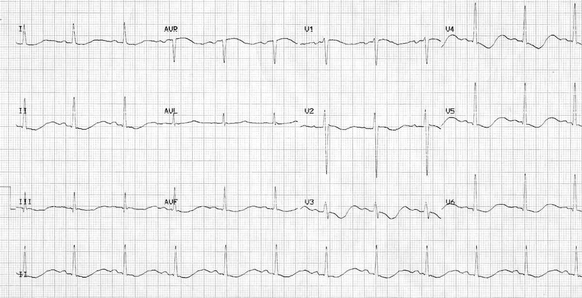 Task for the candidate: You are the pediatric cardiologist. Evaluate this patient, interpret the ECG (U waves, ST depression, prolonged QT), discuss the differential diagnosis of hypokalemia (GI losses, renal losses, transcellular shift, drugs), formulate a diagnostic plan (serum K+, Mg2+, urine electrolytes, ABG), and provide counseling about the causes, management, and monitoring of hypokalemia.
A 10-year-old girl presents to the emergency department with a 1-week history of progressive muscle weakness, fatigue, and constipation. She has had intermittent vomiting over the past 3 days and has been drinking large amounts of water. She is on no regular medications. Her mother reports that she has been tired and has had palpitations on exertion. There is no known heart disease or electrolyte disorder in the family.
Examination: Vital signs: HR 90 bpm (regular), BP 110/70 mm Hg, RR 18/min, SpO2 98% on room air. She is alert but appears lethargic. Neurological examination reveals proximal muscle weakness (difficulty rising from squat) and hyporeflexia. Cardiovascular examination reveals a soft systolic ejection murmur but is otherwise unremarkable. There is no hepatomegaly or edema.
ECG (obtained in ED):
Figure: ECG shows prominent U waves (best seen in V2-V4), ST segment depression, flattened T waves, and prolonged QT interval (QTc 0.50 seconds).
💡 Examiner instruction (interactive): This is a case of Hypokalemia with classic ECG findings. The candidate must recognize the ECG features: prominent U waves (most specific), ST segment depression, flattened T waves, and prolonged QT interval. The candidate should understand the causes of hypokalemia (GI losses: vomiting/diarrhea; renal losses: diuretics, hyperaldosteronism, Bartter/Gitelman; transcellular shift: alkalosis, insulin, hypomagnesemia; drugs: beta-agonists, amphotericin B). Management includes oral or IV potassium replacement, correction of hypomagnesemia if present, and treatment of the underlying cause. The candidate should also discuss the maximum safe IV potassium infusion rate (0.5 mEq/kg/h, max 20 mEq/h) and the importance of cardiac monitoring during IV replacement.
🔍 Examiner Questions (interactive) – Click to reveal model answers
❓ Q1 (Examiner): “Describe the ECG findings in this patient. What are the classic ECG features of hypokalemia?”
✅ Candidate's answer:
• Classic ECG findings in hypokalemia:
1️⃣ Prominent U waves – a small deflection after the T wave, best seen in leads V2-V4. This is the most specific ECG feature of hypokalemia.
2️⃣ ST segment depression – downsloping ST depression.
3️⃣ Flattened T waves – T waves become low amplitude or inverted.
4️⃣ Prolonged QT interval – due to the prolonged repolarization (QTc >0.46 seconds).
5️⃣ Prolonged PR interval – may occur.
6️⃣ Arrhythmias: Premature atrial and ventricular contractions, ventricular tachycardia, and torsades de pointes (especially if QT prolonged).
• Differentiation from hyperkalemia: Hyperkalemia causes peaked (tented) T waves, widening of QRS, and sine wave pattern – not U waves.
• Progression: As hypokalemia worsens, U waves become more prominent, QT prolongation increases, and the risk of arrhythmias rises.
• Classic ECG findings in hypokalemia:
1️⃣ Prominent U waves – a small deflection after the T wave, best seen in leads V2-V4. This is the most specific ECG feature of hypokalemia.
2️⃣ ST segment depression – downsloping ST depression.
3️⃣ Flattened T waves – T waves become low amplitude or inverted.
4️⃣ Prolonged QT interval – due to the prolonged repolarization (QTc >0.46 seconds).
5️⃣ Prolonged PR interval – may occur.
6️⃣ Arrhythmias: Premature atrial and ventricular contractions, ventricular tachycardia, and torsades de pointes (especially if QT prolonged).
• Differentiation from hyperkalemia: Hyperkalemia causes peaked (tented) T waves, widening of QRS, and sine wave pattern – not U waves.
• Progression: As hypokalemia worsens, U waves become more prominent, QT prolongation increases, and the risk of arrhythmias rises.
❓ Q2 (Examiner): “What are the causes of hypokalemia in children? How would you classify them?”
✅ Candidate's answer:
• Causes of hypokalemia:
1️⃣ Gastrointestinal losses: Vomiting (gastric losses), diarrhea, nasogastric suction, laxative abuse.
2️⃣ Renal losses:
- Diuretics: Loop diuretics (furosemide), thiazides.
- Mineralocorticoid excess: Primary hyperaldosteronism (Conn syndrome), secondary hyperaldosteronism (renovascular hypertension, heart failure, cirrhosis), Liddle syndrome (pseudohyperaldosteronism).
- Bartter syndrome: Hypercalciuria, normal Mg, metabolic alkalosis.
- Gitelman syndrome: Hypocalciuria, hypomagnesemia, metabolic alkalosis.
- Renal tubular acidosis (RTA type 1 & 2).
- Drugs: Amphotericin B, aminoglycosides, cisplatin.
3️⃣ Transcellular shift:
- Alkalosis (K+ shifts into cells).
- Insulin/glucose administration.
- Beta-agonists (albuterol).
- Hypokalemic periodic paralysis (SCN4A mutation).
4️⃣ Reduced intake: Malnutrition, anorexia nervosa.
5️⃣ Pseudohypokalemia: Rare – leukocytosis (K+ uptake by WBCs in the specimen).
• Causes of hypokalemia:
1️⃣ Gastrointestinal losses: Vomiting (gastric losses), diarrhea, nasogastric suction, laxative abuse.
2️⃣ Renal losses:
- Diuretics: Loop diuretics (furosemide), thiazides.
- Mineralocorticoid excess: Primary hyperaldosteronism (Conn syndrome), secondary hyperaldosteronism (renovascular hypertension, heart failure, cirrhosis), Liddle syndrome (pseudohyperaldosteronism).
- Bartter syndrome: Hypercalciuria, normal Mg, metabolic alkalosis.
- Gitelman syndrome: Hypocalciuria, hypomagnesemia, metabolic alkalosis.
- Renal tubular acidosis (RTA type 1 & 2).
- Drugs: Amphotericin B, aminoglycosides, cisplatin.
3️⃣ Transcellular shift:
- Alkalosis (K+ shifts into cells).
- Insulin/glucose administration.
- Beta-agonists (albuterol).
- Hypokalemic periodic paralysis (SCN4A mutation).
4️⃣ Reduced intake: Malnutrition, anorexia nervosa.
5️⃣ Pseudohypokalemia: Rare – leukocytosis (K+ uptake by WBCs in the specimen).
❓ Q3 (Examiner): “How do you differentiate Bartter syndrome from Gitelman syndrome? What are the key clinical and laboratory features?”
✅ Candidate's answer:
| Feature | Bartter Syndrome | Gitelman Syndrome |
|---|---|---|
| Onset | Infancy/early childhood | Later childhood/adolescence |
| Hypokalemia | Yes | Yes |
| Metabolic Alkalosis | Yes | Yes |
| Urinary Calcium | Hypercalciuria (↑) | Hypocalciuria (↓) |
| Serum Magnesium | Normal | Hypomagnesemia (↓) |
| BP | Normal (low-normal) | Normal |
| Growth/Development | Growth retardation, polyuria, dehydration | Milder, salt craving, muscle cramps |
| Genetics | NKCC2, ROMK, ClC-Kb | NCC (thiazide-sensitive cotransporter) |
| Treatment | K+ supplements, NSAIDs (indomethacin), spironolactone | K+ and Mg supplements, spironolactone |
❓ Q4 (Examiner): “A child with diabetic ketoacidosis (DKA) has a serum K+ of 5.2 mEq/L. Is total body potassium normal? Why does K+ drop after insulin therapy?”
✅ Candidate's answer:
• Total body potassium in DKA: Depleted despite a normal or high serum K+.
- Mechanism: The hyperglycemia causes osmotic diuresis, leading to urinary potassium losses. Acidosis shifts K+ out of cells into the extracellular space, masking the total body deficit.
• Why K+ drops after insulin:
- Insulin shifts potassium into cells (via Na-K-ATPase).
- As insulin is given, the extracellular K+ falls rapidly, and if not replaced, the patient can develop severe hypokalemia.
• Management: In DKA, start potassium replacement (20-40 mEq/L of IV fluid) once the serum K+ is <5.5 mEq/L and urine output is confirmed. Monitor K+ every 2-4 hours.
• Total body potassium in DKA: Depleted despite a normal or high serum K+.
- Mechanism: The hyperglycemia causes osmotic diuresis, leading to urinary potassium losses. Acidosis shifts K+ out of cells into the extracellular space, masking the total body deficit.
• Why K+ drops after insulin:
- Insulin shifts potassium into cells (via Na-K-ATPase).
- As insulin is given, the extracellular K+ falls rapidly, and if not replaced, the patient can develop severe hypokalemia.
• Management: In DKA, start potassium replacement (20-40 mEq/L of IV fluid) once the serum K+ is <5.5 mEq/L and urine output is confirmed. Monitor K+ every 2-4 hours.
❓ Q5 (Examiner): “A child with hypokalemia (K+ 2.8) and hypomagnesemia (Mg 1.2 mg/dL). After potassium replacement, K+ remains low. Why? What is the management?”
✅ Candidate's answer:
• Mechanism: Magnesium deficiency causes renal potassium wasting. Magnesium is required for the Na-K-ATPase pump and for potassium channel function in the distal tubule. Without adequate magnesium, the kidney cannot retain potassium, leading to refractory hypokalemia.
• Management:
- Correct magnesium deficiency first before aggressively replacing potassium.
- IV magnesium sulfate: 25-50 mg/kg/dose (max 2 g) over 1-2 hours.
- After magnesium is replaced, potassium repletion will be more effective.
- Monitor serum Mg and K+ closely.
- Treat underlying cause of hypomagnesemia (diuretics, Gitelman syndrome, malnutrition).
• Mechanism: Magnesium deficiency causes renal potassium wasting. Magnesium is required for the Na-K-ATPase pump and for potassium channel function in the distal tubule. Without adequate magnesium, the kidney cannot retain potassium, leading to refractory hypokalemia.
• Management:
- Correct magnesium deficiency first before aggressively replacing potassium.
- IV magnesium sulfate: 25-50 mg/kg/dose (max 2 g) over 1-2 hours.
- After magnesium is replaced, potassium repletion will be more effective.
- Monitor serum Mg and K+ closely.
- Treat underlying cause of hypomagnesemia (diuretics, Gitelman syndrome, malnutrition).
❓ Q6 (Examiner): “When would you use IV potassium instead of oral? What is the maximum safe IV infusion rate and concentration?”
✅ Candidate's answer:
• Indications for IV potassium replacement:
- Severe hypokalemia (K+ <3.0 mEq/L) with symptoms (muscle weakness, paralysis, arrhythmias).
- Inability to take oral medications (vomiting, ileus).
- Rapid correction needed (e.g., in DKA, cardiac arrhythmias).
• Maximum safe IV infusion rate:
- 0.5 mEq/kg/h (max 10-20 mEq/h in older children/adults).
- Higher rates (1 mEq/kg/h) require central venous access and continuous cardiac monitoring.
• Maximum concentration (peripheral IV):
- 40 mEq/L (for peripheral line).
- Higher concentrations (up to 80-100 mEq/L) require central venous line.
• Monitoring: Continuous ECG monitoring and frequent serum K+ checks during IV replacement.
• Indications for IV potassium replacement:
- Severe hypokalemia (K+ <3.0 mEq/L) with symptoms (muscle weakness, paralysis, arrhythmias).
- Inability to take oral medications (vomiting, ileus).
- Rapid correction needed (e.g., in DKA, cardiac arrhythmias).
• Maximum safe IV infusion rate:
- 0.5 mEq/kg/h (max 10-20 mEq/h in older children/adults).
- Higher rates (1 mEq/kg/h) require central venous access and continuous cardiac monitoring.
• Maximum concentration (peripheral IV):
- 40 mEq/L (for peripheral line).
- Higher concentrations (up to 80-100 mEq/L) require central venous line.
• Monitoring: Continuous ECG monitoring and frequent serum K+ checks during IV replacement.
❓ Q7 (Examiner): “A 15-year-old presents with acute flaccid paralysis and K+ 2.1 mEq/L. Thyroid function is normal. What is the likely diagnosis and management?”
✅ Candidate's answer:
• Likely diagnosis: Hypokalemic periodic paralysis (primary, autosomal dominant SCN4A mutation) – a channelopathy causing episodic muscle weakness with low K+.
- Triggers: High carbohydrate meals, rest after exercise, emotional stress.
• Differential: Thyrotoxic periodic paralysis (hyperthyroidism), which is more common in Asian males.
• Management:
1️⃣ IV potassium replacement: 0.5 mEq/kg/h (max 20 mEq/h) with ECG monitoring.
2️⃣ Avoid excessive replacement: Rebound hyperkalemia can occur.
3️⃣ Acetazolamide: May be used for prophylaxis.
4️⃣ Genetic counseling: Family screening for SCN4A mutation.
5️⃣ Avoid triggers: High carbohydrate meals, strenuous exercise.
6️⃣ If thyrotoxic: Treat hyperthyroidism.
• Likely diagnosis: Hypokalemic periodic paralysis (primary, autosomal dominant SCN4A mutation) – a channelopathy causing episodic muscle weakness with low K+.
- Triggers: High carbohydrate meals, rest after exercise, emotional stress.
• Differential: Thyrotoxic periodic paralysis (hyperthyroidism), which is more common in Asian males.
• Management:
1️⃣ IV potassium replacement: 0.5 mEq/kg/h (max 20 mEq/h) with ECG monitoring.
2️⃣ Avoid excessive replacement: Rebound hyperkalemia can occur.
3️⃣ Acetazolamide: May be used for prophylaxis.
4️⃣ Genetic counseling: Family screening for SCN4A mutation.
5️⃣ Avoid triggers: High carbohydrate meals, strenuous exercise.
6️⃣ If thyrotoxic: Treat hyperthyroidism.
❓ Q8 (Examiner): “Why is ECG monitoring important during IV potassium replacement? What arrhythmias can occur?”
✅ Candidate's answer:
• ECG monitoring is essential because:
- Potassium has a direct effect on cardiac membrane potential and repolarization.
- Hypokalemia: Delays repolarization (prolonged QT), increases risk of torsades de pointes, ventricular tachycardia, and ventricular fibrillation.
- Overcorrection: Rapid IV potassium can cause hyperkalemia, leading to peaked T waves, wide QRS, and cardiac arrest.
• Arrhythmias associated with hypokalemia:
- Premature atrial contractions (PACs).
- Premature ventricular contractions (PVCs).
- Ventricular tachycardia (VT).
- Torsades de pointes (polymorphic VT with prolonged QT).
- Atrial fibrillation (less common).
• Monitoring: Continuous ECG monitoring during IV potassium replacement, especially if the rate >0.5 mEq/kg/h.
• ECG monitoring is essential because:
- Potassium has a direct effect on cardiac membrane potential and repolarization.
- Hypokalemia: Delays repolarization (prolonged QT), increases risk of torsades de pointes, ventricular tachycardia, and ventricular fibrillation.
- Overcorrection: Rapid IV potassium can cause hyperkalemia, leading to peaked T waves, wide QRS, and cardiac arrest.
• Arrhythmias associated with hypokalemia:
- Premature atrial contractions (PACs).
- Premature ventricular contractions (PVCs).
- Ventricular tachycardia (VT).
- Torsades de pointes (polymorphic VT with prolonged QT).
- Atrial fibrillation (less common).
• Monitoring: Continuous ECG monitoring during IV potassium replacement, especially if the rate >0.5 mEq/kg/h.
❓ Q9 (Examiner): “A 12-year-old with hypertension and hypokalemia (K+ 2.9). Renin is low, aldosterone is high. What is the diagnosis and treatment?”
✅ Candidate's answer:
• Diagnosis: Primary hyperaldosteronism (Conn syndrome) – usually due to an aldosterone-producing adenoma or bilateral adrenal hyperplasia.
- Features: Hypertension + hypokalemia + metabolic alkalosis + suppressed renin + elevated aldosterone (aldosterone:renin ratio >30).
• Management:
1️⃣ Adrenal imaging: CT/MRI to identify adenoma.
2️⃣ Adrenal venous sampling: To lateralize the source.
3️⃣ If adenoma: Surgical resection (adrenalectomy).
4️⃣ If bilateral hyperplasia: Medical therapy with spironolactone (aldosterone antagonist) or eplerenone.
5️⃣ Potassium replacement before surgery.
6️⃣ Avoid: ACE inhibitors (not specific) – spironolactone is first-line.
• Diagnosis: Primary hyperaldosteronism (Conn syndrome) – usually due to an aldosterone-producing adenoma or bilateral adrenal hyperplasia.
- Features: Hypertension + hypokalemia + metabolic alkalosis + suppressed renin + elevated aldosterone (aldosterone:renin ratio >30).
• Management:
1️⃣ Adrenal imaging: CT/MRI to identify adenoma.
2️⃣ Adrenal venous sampling: To lateralize the source.
3️⃣ If adenoma: Surgical resection (adrenalectomy).
4️⃣ If bilateral hyperplasia: Medical therapy with spironolactone (aldosterone antagonist) or eplerenone.
5️⃣ Potassium replacement before surgery.
6️⃣ Avoid: ACE inhibitors (not specific) – spironolactone is first-line.
❓ Q10 (Examiner): “A child with hypertension, hypokalemia, low renin, and low aldosterone. What is the diagnosis and treatment?”
✅ Candidate's answer:
• Diagnosis: Liddle syndrome – a rare autosomal dominant disorder caused by gain-of-function mutations in the epithelial sodium channel (ENaC).
- Features: Early-onset hypertension, hypokalemia, metabolic alkalosis, low renin, low aldosterone (unlike primary hyperaldosteronism).
• Treatment:
- Amiloride or triamterene (ENaC blockers) – first-line.
- Spironolactone is NOT effective (it blocks aldosterone, which is already low).
- Potassium replacement.
- Lifestyle modifications (low-sodium diet).
• Diagnosis: Liddle syndrome – a rare autosomal dominant disorder caused by gain-of-function mutations in the epithelial sodium channel (ENaC).
- Features: Early-onset hypertension, hypokalemia, metabolic alkalosis, low renin, low aldosterone (unlike primary hyperaldosteronism).
• Treatment:
- Amiloride or triamterene (ENaC blockers) – first-line.
- Spironolactone is NOT effective (it blocks aldosterone, which is already low).
- Potassium replacement.
- Lifestyle modifications (low-sodium diet).
❓ Q11 (Examiner): “Which drugs commonly cause hypokalemia in children? What is the mechanism?”
✅ Candidate's answer:
• Drugs causing hypokalemia:
1️⃣ Diuretics: Loop (furosemide) and thiazide diuretics – increase distal tubular K+ excretion.
2️⃣ Corticosteroids: Mineralocorticoid effect → increased K+ excretion.
3️⃣ Beta-agonists: Albuterol (salbutamol) – shifts K+ intracellularly.
4️⃣ Amphotericin B: Renal tubular injury → K+ wasting.
5️⃣ Aminoglycosides: Renal K+ wasting.
6️⃣ Insulin: Shifts K+ into cells.
7️⃣ Laxatives (abuse): GI loss.
8️⃣ Licorice: Glycyrrhizin inhibits 11-beta-hydroxysteroid dehydrogenase → pseudoaldosteronism → hypokalemia.
• Management: Discontinue the offending drug if possible, replace K+, and monitor ECG.
• Drugs causing hypokalemia:
1️⃣ Diuretics: Loop (furosemide) and thiazide diuretics – increase distal tubular K+ excretion.
2️⃣ Corticosteroids: Mineralocorticoid effect → increased K+ excretion.
3️⃣ Beta-agonists: Albuterol (salbutamol) – shifts K+ intracellularly.
4️⃣ Amphotericin B: Renal tubular injury → K+ wasting.
5️⃣ Aminoglycosides: Renal K+ wasting.
6️⃣ Insulin: Shifts K+ into cells.
7️⃣ Laxatives (abuse): GI loss.
8️⃣ Licorice: Glycyrrhizin inhibits 11-beta-hydroxysteroid dehydrogenase → pseudoaldosteronism → hypokalemia.
• Management: Discontinue the offending drug if possible, replace K+, and monitor ECG.
❓ Q12 (Examiner): “A child with hypokalemia and metabolic acidosis. What is the differential diagnosis? Why is this pattern different?”
✅ Candidate's answer:
• Hypokalemia with metabolic acidosis: The differential diagnosis is different from hypokalemia with metabolic alkalosis.
- Causes:
1️⃣ Diarrhea: GI loss of K+ and bicarbonate (alkaline losses).
2️⃣ Renal tubular acidosis (RTA):
- Type 1 (distal RTA): Hypokalemia + metabolic acidosis + hypercalciuria + nephrocalcinosis.
- Type 2 (proximal RTA): Hypokalemia + metabolic acidosis + Fanconi syndrome.
3️⃣ Carbonic anhydrase inhibitors: Acetazolamide.
4️⃣ Amphotericin B: Renal K+ wasting.
5️⃣ Uretero-sigmoidostomy: Rare.
• Key difference: Hypokalemic metabolic acidosis is caused by GI or renal loss of base (bicarbonate) or impaired acid secretion, whereas hypokalemic metabolic alkalosis is caused by loss of acid (vomiting) or mineralocorticoid excess.
• Hypokalemia with metabolic acidosis: The differential diagnosis is different from hypokalemia with metabolic alkalosis.
- Causes:
1️⃣ Diarrhea: GI loss of K+ and bicarbonate (alkaline losses).
2️⃣ Renal tubular acidosis (RTA):
- Type 1 (distal RTA): Hypokalemia + metabolic acidosis + hypercalciuria + nephrocalcinosis.
- Type 2 (proximal RTA): Hypokalemia + metabolic acidosis + Fanconi syndrome.
3️⃣ Carbonic anhydrase inhibitors: Acetazolamide.
4️⃣ Amphotericin B: Renal K+ wasting.
5️⃣ Uretero-sigmoidostomy: Rare.
• Key difference: Hypokalemic metabolic acidosis is caused by GI or renal loss of base (bicarbonate) or impaired acid secretion, whereas hypokalemic metabolic alkalosis is caused by loss of acid (vomiting) or mineralocorticoid excess.
❓ Q13 (Examiner): “A child with chronic kidney disease (CKD) and hypokalemia (K+ 2.8). How would you manage potassium replacement?”
✅ Candidate's answer:
• Management of hypokalemia in CKD:
- Assess urine output: If anuric/oliguric, potassium replacement can be dangerous (risk of hyperkalemia).
- Determine the cause: Renal K+ wasting vs poor intake.
- Oral replacement: Preferred if able to take orally (KCl or K citrate).
- IV replacement: Only if severe symptoms and adequate urine output.
- Monitor K+ frequently: q2-4h.
- Avoid: Potassium-sparing diuretics (spironolactone) if already hyperkalemic.
- Treat underlying cause: If due to diuretics, adjust dose or switch to K+-sparing diuretic.
- Dietary counseling: Increase K+ rich foods (if not contraindicated).
• Management of hypokalemia in CKD:
- Assess urine output: If anuric/oliguric, potassium replacement can be dangerous (risk of hyperkalemia).
- Determine the cause: Renal K+ wasting vs poor intake.
- Oral replacement: Preferred if able to take orally (KCl or K citrate).
- IV replacement: Only if severe symptoms and adequate urine output.
- Monitor K+ frequently: q2-4h.
- Avoid: Potassium-sparing diuretics (spironolactone) if already hyperkalemic.
- Treat underlying cause: If due to diuretics, adjust dose or switch to K+-sparing diuretic.
- Dietary counseling: Increase K+ rich foods (if not contraindicated).
❓ Q14 (Examiner): “The parents are worried about their child's condition. How will you counsel them?”
✅ Candidate's structured answer:
• “Your child has a condition called hypokalemia – a low level of potassium in the blood. Potassium is very important for muscle function, including the heart muscle.”
• “The ECG shows some characteristic changes – U waves, ST depression, and a prolonged QT interval – which are signs of low potassium. This can cause muscle weakness and, if severe, heart rhythm problems.”
• “The good news is that this is very treatable. We will give your child potassium supplements – either by mouth or through an IV, depending on the severity. We will monitor the heart rhythm closely to make sure it is safe.”
• “We will also find out why the potassium is low – it could be from vomiting, certain medications, or an underlying kidney condition. Once we find the cause, we can treat it and prevent it from happening again.”
• “With proper treatment, your child should recover completely. We will keep you updated every step of the way.”
• “Your child has a condition called hypokalemia – a low level of potassium in the blood. Potassium is very important for muscle function, including the heart muscle.”
• “The ECG shows some characteristic changes – U waves, ST depression, and a prolonged QT interval – which are signs of low potassium. This can cause muscle weakness and, if severe, heart rhythm problems.”
• “The good news is that this is very treatable. We will give your child potassium supplements – either by mouth or through an IV, depending on the severity. We will monitor the heart rhythm closely to make sure it is safe.”
• “We will also find out why the potassium is low – it could be from vomiting, certain medications, or an underlying kidney condition. Once we find the cause, we can treat it and prevent it from happening again.”
• “With proper treatment, your child should recover completely. We will keep you updated every step of the way.”
🗣️ Examiner's probing / high-yield points (ECG of Hypokalemia):
• "What are the classic ECG findings in hypokalemia?" → U waves, ST depression, flattened T waves, prolonged QT.
• "What is the most common cause of hypokalemia in children?" → GI losses (vomiting, diarrhea) and diuretics.
• "What is the difference between Bartter and Gitelman syndrome?" → Bartter: hypercalciuria, normal Mg. Gitelman: hypocalciuria, hypomagnesemia.
• "Why does hypomagnesemia cause refractory hypokalemia?" → Mg deficiency impairs renal K+ retention.
• "What is the maximum safe IV K+ rate?" → 0.5 mEq/kg/h (max 20 mEq/h).
• "What is the treatment for Liddle syndrome?" → Amiloride (ENaC blocker).
• "What is the treatment for primary hyperaldosteronism?" → Spironolactone or adrenalectomy.
• "What is the ECG finding in hyperkalemia?" → Peaked T waves, wide QRS, sine wave.
• "What are the classic ECG findings in hypokalemia?" → U waves, ST depression, flattened T waves, prolonged QT.
• "What is the most common cause of hypokalemia in children?" → GI losses (vomiting, diarrhea) and diuretics.
• "What is the difference between Bartter and Gitelman syndrome?" → Bartter: hypercalciuria, normal Mg. Gitelman: hypocalciuria, hypomagnesemia.
• "Why does hypomagnesemia cause refractory hypokalemia?" → Mg deficiency impairs renal K+ retention.
• "What is the maximum safe IV K+ rate?" → 0.5 mEq/kg/h (max 20 mEq/h).
• "What is the treatment for Liddle syndrome?" → Amiloride (ENaC blocker).
• "What is the treatment for primary hyperaldosteronism?" → Spironolactone or adrenalectomy.
• "What is the ECG finding in hyperkalemia?" → Peaked T waves, wide QRS, sine wave.
📘 ECG of Hypokalemia – Core Revision for TOACS
⚡ Definition
Hypokalemia: Serum K+ <3.5 mEq/L. Causes: GI losses, renal losses, transcellular shift, drugs, reduced intake.
Hypokalemia: Serum K+ <3.5 mEq/L. Causes: GI losses, renal losses, transcellular shift, drugs, reduced intake.
📊 ECG Findings
Prominent U waves (V2-V4), ST depression, flattened T waves, prolonged QT, arrhythmias (PACs, PVCs, VT, torsades).
Prominent U waves (V2-V4), ST depression, flattened T waves, prolonged QT, arrhythmias (PACs, PVCs, VT, torsades).
🔍 Causes
GI: vomiting, diarrhea. Renal: diuretics, hyperaldosteronism, Bartter/Gitelman, RTA, drugs (amphotericin B). Transcellular: alkalosis, insulin, beta-agonists.
GI: vomiting, diarrhea. Renal: diuretics, hyperaldosteronism, Bartter/Gitelman, RTA, drugs (amphotericin B). Transcellular: alkalosis, insulin, beta-agonists.
💊 Management
Oral KCl (safe). IV KCl: 0.5 mEq/kg/h (max 20 mEq/h), concentration 40 mEq/L (peripheral). Correct Mg first if hypomagnesemia.
Oral KCl (safe). IV KCl: 0.5 mEq/kg/h (max 20 mEq/h), concentration 40 mEq/L (peripheral). Correct Mg first if hypomagnesemia.
🧬 Bartter vs Gitelman
Bartter: hypercalciuria, normal Mg, infancy. Gitelman: hypocalciuria, hypomagnesemia, later onset.
Bartter: hypercalciuria, normal Mg, infancy. Gitelman: hypocalciuria, hypomagnesemia, later onset.
⚠️ Complications
Muscle weakness, paralysis, rhabdomyolysis, arrhythmias (torsades de pointes), ileus, polyuria.
Muscle weakness, paralysis, rhabdomyolysis, arrhythmias (torsades de pointes), ileus, polyuria.
⭐ High-yield pearls for TOACS (ECG of Hypokalemia):
• ECG: U waves + ST depression + prolonged QT.
• Most common cause: GI losses and diuretics.
• Bartter vs Gitelman: Hypercalciuria vs hypocalciuria.
• Treat hypomagnesemia first if present.
• IV K+: 0.5 mEq/kg/h max, 40 mEq/L peripheral.
• Liddle syndrome: Amiloride (ENaC blocker).
• Primary hyperaldosteronism: Spironolactone.
• ECG: U waves + ST depression + prolonged QT.
• Most common cause: GI losses and diuretics.
• Bartter vs Gitelman: Hypercalciuria vs hypocalciuria.
• Treat hypomagnesemia first if present.
• IV K+: 0.5 mEq/kg/h max, 40 mEq/L peripheral.
• Liddle syndrome: Amiloride (ENaC blocker).
• Primary hyperaldosteronism: Spironolactone.
🗣️ Candidate's role-play & examiner feedback
💬 To the candidate (role‑play): You will be asked the 14 questions from the Examiner Q&A tab. This station tests knowledge of ECG of Hypokalemia – classic ECG findings (U waves, ST depression, prolonged QT), causes (GI losses, renal losses, transcellular shift), differential diagnosis (Bartter vs Gitelman, Liddle, hyperaldosteronism), management (IV/oral K+ replacement, treat hypomagnesemia), and complications (arrhythmias). Provide compassionate counseling to parents about the condition and its treatment.
📝 Examiner Marking Grid (ECG of Hypokalemia – TOACS station):
- ✅ Interprets ECG: U waves, ST depression, flattened T waves, prolonged QT
- ✅ Lists causes: GI losses, renal losses (diuretics, hyperaldosteronism, Bartter/Gitelman), transcellular shift
- ✅ Differentiates Bartter (hypercalciuria) from Gitelman (hypocalciuria, hypomagnesemia)
- ✅ Manages IV K+ replacement: 0.5 mEq/kg/h (max 20 mEq/h), 40 mEq/L peripheral
- ✅ States role of ECG monitoring and arrhythmia risks
- ✅ Discusses hypomagnesemia and refractory hypokalemia
- ✅ Diagnoses Liddle syndrome (low renin, low aldosterone, amiloride)
- ✅ Diagnoses primary hyperaldosteronism (high aldosterone, spironolactone)
- ✅ Provides compassionate counseling and discusses prognosis
📚 Key references: Nelson Textbook of Pediatrics 22e (Section 73.4 – Potassium Disorders), Pediatric Electrocardiography guidelines.