FCPS Paediatrics TOACS · Interactive Station
🩺 Cephalohematoma — Subperiosteal hemorrhage, scalp swelling, benign course, complications
📚 Paeds Online – paeds.online
⚕️ OBSERVED STATION · CPSP FORMAT · 8 MINUTES · SEPARATE TABS · CLINICAL PHOTO INCLUDED
📖 Problem-oriented Clinical Scenario + Photograph
👶🏻 Clinical Scenario (read aloud – 2 min):
A 24-hour-old term male infant is evaluated in the newborn nursery. He was born at 40 weeks via vacuum-assisted vaginal delivery due to prolonged second stage. The mother had an uncomplicated pregnancy. Birth weight 3.8 kg. On examination: The infant is well-appearing, feeding well, and vital signs are normal. A firm, non-pulsatile, fluctuant swelling measuring approximately 5×4 cm is noted over the right parietal region. The swelling does not cross the sagittal suture line. The overlying skin is normal, with no bruising or abrasion. The anterior fontanelle is soft and flat. Neurological examination is normal. The mother is concerned about the swelling and asks what it is and whether it will affect the baby's brain.
A clinical photograph of the scalp swelling is shown below.
Task: Describe the findings, propose the most likely diagnosis, differentiate from other scalp swellings (caput succedaneum, subgaleal hemorrhage), discuss management, complications (hyperbilirubinemia, anemia), and parental counseling.
A 24-hour-old term male infant is evaluated in the newborn nursery. He was born at 40 weeks via vacuum-assisted vaginal delivery due to prolonged second stage. The mother had an uncomplicated pregnancy. Birth weight 3.8 kg. On examination: The infant is well-appearing, feeding well, and vital signs are normal. A firm, non-pulsatile, fluctuant swelling measuring approximately 5×4 cm is noted over the right parietal region. The swelling does not cross the sagittal suture line. The overlying skin is normal, with no bruising or abrasion. The anterior fontanelle is soft and flat. Neurological examination is normal. The mother is concerned about the swelling and asks what it is and whether it will affect the baby's brain.
A clinical photograph of the scalp swelling is shown below.
Task: Describe the findings, propose the most likely diagnosis, differentiate from other scalp swellings (caput succedaneum, subgaleal hemorrhage), discuss management, complications (hyperbilirubinemia, anemia), and parental counseling.
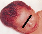
🔍 Figure: Right parietal cephalohematoma – a firm, fluctuant, well-defined swelling limited to one cranial bone (does not cross the sagittal suture). Overlying skin is intact. This is a subperiosteal hemorrhage, typically following vacuum- or forceps-assisted delivery.
💡 Examiner instruction (interactive): The candidate will be asked to identify cephalohematoma, differentiate from caput succedaneum and subgaleal hemorrhage, discuss etiology (subperiosteal bleeding), management (observation, no intervention), complications (hyperbilirubinemia, anemia, rare osteomyelitis), and provide parental counseling (benign, spontaneous resolution, reassurance).
🔍 Examiner Questions (interactive) – Click to reveal model answers
❓ Q1 (Examiner): “Describe the findings in the image. What is the most likely diagnosis?”
✅ Candidate's structured answer:
• Findings: Well-defined, firm, fluctuant, non-pulsatile swelling over the right parietal bone. Does NOT cross the sagittal suture line. Overlying skin is normal, no erythema or abrasion.
• Diagnosis: Cephalohematoma – a subperiosteal hemorrhage typically associated with vacuum-assisted or forceps delivery.
• Findings: Well-defined, firm, fluctuant, non-pulsatile swelling over the right parietal bone. Does NOT cross the sagittal suture line. Overlying skin is normal, no erythema or abrasion.
• Diagnosis: Cephalohematoma – a subperiosteal hemorrhage typically associated with vacuum-assisted or forceps delivery.
❓ Q2 (Examiner): “What is the underlying pathology of cephalohematoma? Why does it not cross suture lines?”
✅ Candidate's answer:
• Pathology: Bleeding into the subperiosteal space (between the skull periosteum and the calvarial bone) due to rupture of small emissary veins or diploic veins during birth trauma.
• The periosteum is tightly adherent to the skull bone at the suture lines (sutures are periosteal reflections). Therefore the hematoma is confined to a single cranial bone and does not cross sutures.
• Risk factors: Vacuum extraction (most common), forceps delivery, prolonged labor, macrosomia, primiparity.
• Incidence: 1-2% of all live births, higher with instrumental deliveries (up to 10-15% with vacuum).
• Pathology: Bleeding into the subperiosteal space (between the skull periosteum and the calvarial bone) due to rupture of small emissary veins or diploic veins during birth trauma.
• The periosteum is tightly adherent to the skull bone at the suture lines (sutures are periosteal reflections). Therefore the hematoma is confined to a single cranial bone and does not cross sutures.
• Risk factors: Vacuum extraction (most common), forceps delivery, prolonged labor, macrosomia, primiparity.
• Incidence: 1-2% of all live births, higher with instrumental deliveries (up to 10-15% with vacuum).
❓ Q3 (Examiner): “What are the differential diagnoses of a neonatal scalp swelling? Differentiate from caput succedaneum and subgaleal hemorrhage.”
✅ Candidate's answer:
• Cephalohematoma: Subperiosteal, confined to one bone, does not cross sutures, firm/fluctuant, appears hours to days after birth, resolves over weeks to months.
• Caput succedaneum: Subcutaneous (edema), crosses suture lines, soft/pitting, present at birth, resolves within days (24-72 hours). Associated with vertex delivery.
• Subgaleal hemorrhage: Subaponeurotic (potential space), crosses sutures, fluctuant (can spread to entire scalp), may be massive (up to 40% blood volume), presents with hypovolemic shock, requires urgent intervention. Associated with vacuum extraction.
• Other: Meningocele/en-cephalocele (midline, pulsatile, transilluminates), skull fracture (may underlie cephalohematoma in 10-25%).
• Cephalohematoma: Subperiosteal, confined to one bone, does not cross sutures, firm/fluctuant, appears hours to days after birth, resolves over weeks to months.
• Caput succedaneum: Subcutaneous (edema), crosses suture lines, soft/pitting, present at birth, resolves within days (24-72 hours). Associated with vertex delivery.
• Subgaleal hemorrhage: Subaponeurotic (potential space), crosses sutures, fluctuant (can spread to entire scalp), may be massive (up to 40% blood volume), presents with hypovolemic shock, requires urgent intervention. Associated with vacuum extraction.
• Other: Meningocele/en-cephalocele (midline, pulsatile, transilluminates), skull fracture (may underlie cephalohematoma in 10-25%).
❓ Q4 (Examiner): “What is the natural history of cephalohematoma? How long does it take to resolve?”
✅ Candidate's answer:
• Spontaneous resolution occurs in most cases without intervention.
• Timeline:
- Swelling may increase in size over first 24-48 hours (continued slow bleeding).
- Firmness increases over first week.
- Gradual resorption begins by 2-3 weeks.
- Complete resolution typically by 2-4 months (sometimes up to 6 months).
- May leave a calcified rim (residual bony ridge) that resolves over months to years (sometimes persists as a palpable thickening).
• No treatment needed. Avoid aspiration – risk of infection (osteomyelitis).
• Spontaneous resolution occurs in most cases without intervention.
• Timeline:
- Swelling may increase in size over first 24-48 hours (continued slow bleeding).
- Firmness increases over first week.
- Gradual resorption begins by 2-3 weeks.
- Complete resolution typically by 2-4 months (sometimes up to 6 months).
- May leave a calcified rim (residual bony ridge) that resolves over months to years (sometimes persists as a palpable thickening).
• No treatment needed. Avoid aspiration – risk of infection (osteomyelitis).
❓ Q5 (Examiner): “What are the potential complications of cephalohematoma?”
✅ Candidate's answer:
• Hyperbilirubinemia (most common complication, ~20-30%): Breakdown of hematoma releases hemoglobin → increased bilirubin load → exaggerated physiologic jaundice or pathological jaundice requiring phototherapy.
• Anemia (rare, large hematomas): Significant blood sequestration (up to 50-100 mL) can cause anemia requiring transfusion – very rare.
• Infection (very rare): Osteomyelitis of the underlying skull bone (if aspiration attempted or spontaneous infection). Presents with erythema, warmth, tenderness, fever. Organisms: S. aureus, E. coli.
• Calcification / ossification: May leave a "bony knob" that persists for years but resolves eventually.
• Associated skull fracture: Linear fracture in 10-25% of cases – usually incidental, no treatment needed.
• Discomfort: Generally painless; infant usually asymptomatic.
• Hyperbilirubinemia (most common complication, ~20-30%): Breakdown of hematoma releases hemoglobin → increased bilirubin load → exaggerated physiologic jaundice or pathological jaundice requiring phototherapy.
• Anemia (rare, large hematomas): Significant blood sequestration (up to 50-100 mL) can cause anemia requiring transfusion – very rare.
• Infection (very rare): Osteomyelitis of the underlying skull bone (if aspiration attempted or spontaneous infection). Presents with erythema, warmth, tenderness, fever. Organisms: S. aureus, E. coli.
• Calcification / ossification: May leave a "bony knob" that persists for years but resolves eventually.
• Associated skull fracture: Linear fracture in 10-25% of cases – usually incidental, no treatment needed.
• Discomfort: Generally painless; infant usually asymptomatic.
❓ Q6 (Examiner): “How does cephalohematoma contribute to hyperbilirubinemia? How do you monitor and manage it?”
✅ Candidate's answer:
• Mechanism: Breakdown of extravasated red blood cells from the hematoma increases the bilirubin load on the neonatal liver (heme oxygenase pathway). The immature liver cannot conjugate the increased bilirubin efficiently → unconjugated hyperbilirubinemia.
• Monitoring:
- Serial bilirubin measurements (transcutaneous or serum) every 12-24 hours for first 3-5 days, especially if large hematoma.
- Monitor for jaundice progression (cephalocaudal spread).
• Management:
- Ensure adequate hydration and frequent feeds.
- Phototherapy if bilirubin exceeds age-specific nomogram thresholds (see AAP guidelines).
- Exchange transfusion rarely needed.
• Prevention: No specific prevention; awareness of increased risk.
• Mechanism: Breakdown of extravasated red blood cells from the hematoma increases the bilirubin load on the neonatal liver (heme oxygenase pathway). The immature liver cannot conjugate the increased bilirubin efficiently → unconjugated hyperbilirubinemia.
• Monitoring:
- Serial bilirubin measurements (transcutaneous or serum) every 12-24 hours for first 3-5 days, especially if large hematoma.
- Monitor for jaundice progression (cephalocaudal spread).
• Management:
- Ensure adequate hydration and frequent feeds.
- Phototherapy if bilirubin exceeds age-specific nomogram thresholds (see AAP guidelines).
- Exchange transfusion rarely needed.
• Prevention: No specific prevention; awareness of increased risk.
❓ Q7 (Examiner): “Can a cephalohematoma cause significant anemia? When should you suspect it?”
✅ Candidate's answer:
• Yes, but rare. Very large cephalohematomas can sequester 50-100 mL of blood (total neonatal blood volume ~80-100 mL/kg; for 3.5 kg infant ≈ 300 mL). Significant hemorrhage into the subperiosteal space can cause hypovolemia and anemia.
• Suspect anemia if:
- Pale appearance, poor feeding, lethargy, tachycardia, tachypnea.
- Hematoma is very large (>6-8 cm diameter).
- Fall in hemoglobin/hematocrit on serial CBC.
• Management:
- Monitor hemoglobin.
- If symptomatic and Hb < 10 g/dL → packed red blood cell transfusion.
- Iron supplementation after resolution to replenish iron stores.
• Most cephalohematomas do not cause clinically significant anemia.
• Yes, but rare. Very large cephalohematomas can sequester 50-100 mL of blood (total neonatal blood volume ~80-100 mL/kg; for 3.5 kg infant ≈ 300 mL). Significant hemorrhage into the subperiosteal space can cause hypovolemia and anemia.
• Suspect anemia if:
- Pale appearance, poor feeding, lethargy, tachycardia, tachypnea.
- Hematoma is very large (>6-8 cm diameter).
- Fall in hemoglobin/hematocrit on serial CBC.
• Management:
- Monitor hemoglobin.
- If symptomatic and Hb < 10 g/dL → packed red blood cell transfusion.
- Iron supplementation after resolution to replenish iron stores.
• Most cephalohematomas do not cause clinically significant anemia.
❓ Q8 (Examiner): “What is the risk of osteomyelitis with cephalohematoma? How does it present and how is it treated?”
✅ Candidate's answer:
• Risk: Extremely low (<1%) unless aspiration or needle drainage is attempted (then risk increases significantly). Spontaneous infection is rare.
• Presentation (usually 2-6 weeks of age):
- Swelling becomes erythematous, warm, tender (previously non-tender).
- Overlying skin may become tense, shiny.
- Fever, irritability, poor feeding, elevated inflammatory markers (CRP, WBC).
- Associated with skull osteomyelitis (underlying bone infection).
• Pathogens: S. aureus, Group B Strep, E. coli, Salmonella (rare).
• Management:
- IV antibiotics (vancomycin + cefotaxime or cloxacillin + gentamicin).
- Imaging: Ultrasound or MRI to evaluate for abscess or bone involvement.
- Surgical drainage if abscess present.
- Antibiotics for 3-6 weeks depending on severity.
• Prevention: Do NOT aspirate or drain uninfected cephalohematoma.
• Risk: Extremely low (<1%) unless aspiration or needle drainage is attempted (then risk increases significantly). Spontaneous infection is rare.
• Presentation (usually 2-6 weeks of age):
- Swelling becomes erythematous, warm, tender (previously non-tender).
- Overlying skin may become tense, shiny.
- Fever, irritability, poor feeding, elevated inflammatory markers (CRP, WBC).
- Associated with skull osteomyelitis (underlying bone infection).
• Pathogens: S. aureus, Group B Strep, E. coli, Salmonella (rare).
• Management:
- IV antibiotics (vancomycin + cefotaxime or cloxacillin + gentamicin).
- Imaging: Ultrasound or MRI to evaluate for abscess or bone involvement.
- Surgical drainage if abscess present.
- Antibiotics for 3-6 weeks depending on severity.
• Prevention: Do NOT aspirate or drain uninfected cephalohematoma.
❓ Q9 (Examiner): “When is imaging (skull X-ray, CT, ultrasound) indicated for a cephalohematoma?”
✅ Candidate's answer:
• Routine imaging is NOT indicated for typical, uncomplicated cephalohematoma. Diagnosis is clinical.
• Indications for skull X-ray:
- Suspected underlying depressed skull fracture (palpable depression, focal bony step-off).
- Associated neurological symptoms (seizures, altered sensorium).
- Large, expanding cephalohematoma beyond 48 hours.
• Indications for ultrasound or CT:
- Suspected subgaleal hemorrhage (if swelling crosses sutures rapidly).
- Suspected infection (abscess, osteomyelitis) – ultrasound to detect fluid collection, CT/MRI for bone involvement.
- Neurological symptoms (intracranial bleed) – CT or MRI.
• Finding: Skull X-ray may show linear skull fracture underlying the cephalohematoma in 10-25% of cases – usually no intervention needed.
• Routine imaging is NOT indicated for typical, uncomplicated cephalohematoma. Diagnosis is clinical.
• Indications for skull X-ray:
- Suspected underlying depressed skull fracture (palpable depression, focal bony step-off).
- Associated neurological symptoms (seizures, altered sensorium).
- Large, expanding cephalohematoma beyond 48 hours.
• Indications for ultrasound or CT:
- Suspected subgaleal hemorrhage (if swelling crosses sutures rapidly).
- Suspected infection (abscess, osteomyelitis) – ultrasound to detect fluid collection, CT/MRI for bone involvement.
- Neurological symptoms (intracranial bleed) – CT or MRI.
• Finding: Skull X-ray may show linear skull fracture underlying the cephalohematoma in 10-25% of cases – usually no intervention needed.
❓ Q10 (Examiner): “What is the association between cephalohematoma and skull fracture? How is it managed?”
✅ Candidate's answer:
• Incidence: Underlying linear skull fracture occurs in 10-25% of cephalohematomas (especially after forceps delivery).
• Clinical presentation: Usually asymptomatic; fracture is detected only if X-ray or CT is obtained.
• Management:
- Linear, non-depressed fractures require no treatment – heal spontaneously in weeks to months.
- Depressed ("ping-pong") fractures: Rare; may require surgical elevation if neurological deficit or cosmetic concerns.
- Monitor for complications: leptomeningeal cyst (growing skull fracture) – extremely rare; presents as persistent, enlarging pulsatile swelling months later.
• Clinical pearl: Do not routinely image every cephalohematoma; only if neurological signs, depressed fracture palpable, or persistent.
• Incidence: Underlying linear skull fracture occurs in 10-25% of cephalohematomas (especially after forceps delivery).
• Clinical presentation: Usually asymptomatic; fracture is detected only if X-ray or CT is obtained.
• Management:
- Linear, non-depressed fractures require no treatment – heal spontaneously in weeks to months.
- Depressed ("ping-pong") fractures: Rare; may require surgical elevation if neurological deficit or cosmetic concerns.
- Monitor for complications: leptomeningeal cyst (growing skull fracture) – extremely rare; presents as persistent, enlarging pulsatile swelling months later.
• Clinical pearl: Do not routinely image every cephalohematoma; only if neurological signs, depressed fracture palpable, or persistent.
❓ Q11 (Examiner): “How will you manage this infant? What will you tell the anxious mother?”
✅ Candidate's structured answer:
• Management: Observation, NO intervention.
• Parental counseling points:
1️⃣ “This is a cephalohematoma – a collection of blood under the outer covering of the skull bone. It is NOT inside the brain and does NOT affect the brain.”
2️⃣ “It will go away completely on its own over 2-4 months. No treatment or medicine is needed.”
3️⃣ “Do NOT apply anything to it, do NOT massage it, and do NOT try to drain it. Squeezing or aspiration can cause serious infection.”
4️⃣ “It may feel firm or hard at first, then get softer over time. It might leave a small bump (calcified rim) that will also go away.”
5️⃣ “We will monitor your baby for jaundice (yellow skin) because breaking down the blood can cause higher bilirubin levels.”
6️⃣ “If the swelling becomes red, warm, tender, or your baby develops fever, come back immediately – that could be a sign of infection.”
7️⃣ “You can hold, feed, and bathe the baby normally. The hematoma is not painful.”
• Management: Observation, NO intervention.
• Parental counseling points:
1️⃣ “This is a cephalohematoma – a collection of blood under the outer covering of the skull bone. It is NOT inside the brain and does NOT affect the brain.”
2️⃣ “It will go away completely on its own over 2-4 months. No treatment or medicine is needed.”
3️⃣ “Do NOT apply anything to it, do NOT massage it, and do NOT try to drain it. Squeezing or aspiration can cause serious infection.”
4️⃣ “It may feel firm or hard at first, then get softer over time. It might leave a small bump (calcified rim) that will also go away.”
5️⃣ “We will monitor your baby for jaundice (yellow skin) because breaking down the blood can cause higher bilirubin levels.”
6️⃣ “If the swelling becomes red, warm, tender, or your baby develops fever, come back immediately – that could be a sign of infection.”
7️⃣ “You can hold, feed, and bathe the baby normally. The hematoma is not painful.”
❓ Q12 (Examiner): “What follow-up is required? When would you refer to a neurosurgeon or pediatric surgeon?”
✅ Candidate's answer:
• Follow-up:
- Routine newborn care; re-examine at 2-week and 2-month visits.
- Monitor bilirubin levels for first 5 days (if hematoma large).
- Document size and resolution over time.
• Referral indications (to pediatric neurosurgeon or surgery):
1️⃣ Depressed skull fracture with neurological deficit.
2️⃣ Growing skull fracture (leptomeningeal cyst) – rare: persistent, enlarging, pulsatile swelling.
3️⃣ Suspected osteomyelitis or abscess (red, tender, fluctuant with fever).
4️⃣ Cosmetically concerning persistent calcified ridge after 1 year of age (rare).
• Most cephalohematomas do NOT require subspecialty referral.
• Follow-up:
- Routine newborn care; re-examine at 2-week and 2-month visits.
- Monitor bilirubin levels for first 5 days (if hematoma large).
- Document size and resolution over time.
• Referral indications (to pediatric neurosurgeon or surgery):
1️⃣ Depressed skull fracture with neurological deficit.
2️⃣ Growing skull fracture (leptomeningeal cyst) – rare: persistent, enlarging, pulsatile swelling.
3️⃣ Suspected osteomyelitis or abscess (red, tender, fluctuant with fever).
4️⃣ Cosmetically concerning persistent calcified ridge after 1 year of age (rare).
• Most cephalohematomas do NOT require subspecialty referral.
❓ Q13 (Examiner): “How can you reduce the risk of significant hyperbilirubinemia in an infant with a large cephalohematoma?”
✅ Candidate's answer:
• Cannot prevent completely – the hematoma will resolve and release bilirubin.
• Minimize severity by:
1️⃣ Adequate hydration and feeding: Frequent breastfeeding (8-12 times/day) or formula feeding to promote stooling (excretion of bilirubin).
2️⃣ Avoid dehydration: Monitor weight loss (<10% of birth weight).
3️⃣ Early recognition: Perform bilirubin screening at 24-48 hours (transcutaneous or serum).
4️⃣ Early phototherapy if bilirubin reaches age-specific threshold (do not wait for dangerous levels).
5️⃣ Monitor for hyperbilirubinemia risk factors: Prematurity, poor feeding, ABO/Rh incompatibility.
• No role for prophylactic phototherapy or medications.
• Cannot prevent completely – the hematoma will resolve and release bilirubin.
• Minimize severity by:
1️⃣ Adequate hydration and feeding: Frequent breastfeeding (8-12 times/day) or formula feeding to promote stooling (excretion of bilirubin).
2️⃣ Avoid dehydration: Monitor weight loss (<10% of birth weight).
3️⃣ Early recognition: Perform bilirubin screening at 24-48 hours (transcutaneous or serum).
4️⃣ Early phototherapy if bilirubin reaches age-specific threshold (do not wait for dangerous levels).
5️⃣ Monitor for hyperbilirubinemia risk factors: Prematurity, poor feeding, ABO/Rh incompatibility.
• No role for prophylactic phototherapy or medications.
❓ Q14 (Examiner): “What is the long-term prognosis for this infant? Are there any lasting effects?”
✅ Candidate's answer:
• Excellent prognosis. Complete resolution expected.
• No long-term effects on:
- Brain development or neurological function.
- Skull growth or shape.
- Head circumference.
- Cognitive abilities.
• Possible temporary findings (not sequelae):
- Palpable calcified rim (osseous ridge) that resolves by 1-2 years of age.
- Mild cosmetic irregularity (usually unnoticeable).
• No increased risk of future intracranial bleeding or seizures.
• Final reassurance to parents: “Your baby will recover completely, and the swelling will disappear over the next few months. No treatment needed.”
• Excellent prognosis. Complete resolution expected.
• No long-term effects on:
- Brain development or neurological function.
- Skull growth or shape.
- Head circumference.
- Cognitive abilities.
• Possible temporary findings (not sequelae):
- Palpable calcified rim (osseous ridge) that resolves by 1-2 years of age.
- Mild cosmetic irregularity (usually unnoticeable).
• No increased risk of future intracranial bleeding or seizures.
• Final reassurance to parents: “Your baby will recover completely, and the swelling will disappear over the next few months. No treatment needed.”
🗣️ Examiner's probing / high-yield points:
• "What is the key distinguishing feature from caput succedaneum?" → Cephalohematoma does NOT cross sutures; caput crosses sutures.
• "What is the most common complication?" → Hyperbilirubinemia (due to breakdown of blood).
• "Why should you never aspirate a cephalohematoma?" → Risk of introducing infection → osteomyelitis.
• "What is subgaleal hemorrhage?" → Bleeding into subaponeurotic space; crosses sutures, can cause shock. Emergency.
• "What percentage have underlying skull fracture?" → 10-25%.
• "When does cephalohematoma resolve?" → 2-4 months (calcified rim may persist longer).
• "What is the most important parental advice?" → Do NOT apply anything or try to drain it; monitor for jaundice.
• "What is the key distinguishing feature from caput succedaneum?" → Cephalohematoma does NOT cross sutures; caput crosses sutures.
• "What is the most common complication?" → Hyperbilirubinemia (due to breakdown of blood).
• "Why should you never aspirate a cephalohematoma?" → Risk of introducing infection → osteomyelitis.
• "What is subgaleal hemorrhage?" → Bleeding into subaponeurotic space; crosses sutures, can cause shock. Emergency.
• "What percentage have underlying skull fracture?" → 10-25%.
• "When does cephalohematoma resolve?" → 2-4 months (calcified rim may persist longer).
• "What is the most important parental advice?" → Do NOT apply anything or try to drain it; monitor for jaundice.
📘 Cephalohematoma – Core Revision for TOACS
🔍 Definition
Subperiosteal hemorrhage confined to a single cranial bone. Does NOT cross suture lines. Incidence 1-2% of live births, higher with vacuum/forceps.
Subperiosteal hemorrhage confined to a single cranial bone. Does NOT cross suture lines. Incidence 1-2% of live births, higher with vacuum/forceps.
📊 Clinical Features
Firm, fluctuant, non-pulsatile swelling appearing hours to days after birth. Overlying skin normal. Resolves over 2-4 months.
Firm, fluctuant, non-pulsatile swelling appearing hours to days after birth. Overlying skin normal. Resolves over 2-4 months.
🩺 Differential Diagnosis
Caput succedaneum (crosses sutures, resolves days), subgaleal hemorrhage (crosses sutures, massive, shock).
Caput succedaneum (crosses sutures, resolves days), subgaleal hemorrhage (crosses sutures, massive, shock).
⚕️ Management
Observation only. NO aspiration, NO massage, NO compression. Manage hyperbilirubinemia if develops.
Observation only. NO aspiration, NO massage, NO compression. Manage hyperbilirubinemia if develops.
⚠️ Complications
Hyperbilirubinemia (20-30%), anemia (rare), infection/osteomyelitis (very rare if not aspirated), associated linear skull fracture (10-25%, incidental).
Hyperbilirubinemia (20-30%), anemia (rare), infection/osteomyelitis (very rare if not aspirated), associated linear skull fracture (10-25%, incidental).
📈 Prognosis
Excellent. Complete resolution. No long-term neurological or cosmetic sequelae.
Excellent. Complete resolution. No long-term neurological or cosmetic sequelae.
⭐ High-yield pearls for TOACS (Cephalohematoma):
• Clinical hallmark: Swelling confined to one cranial bone, does NOT cross suture lines.
• Most common complication: Hyperbilirubinemia – monitor bilirubin.
• Do NOT aspirate – risk of osteomyelitis.
• Differentiate from subgaleal hemorrhage: Subgaleal crosses sutures, fluctuant, may cause shock.
• Natural history: Resolves in 2-4 months; may leave a calcified rim.
• Parental reassurance: Does NOT affect the brain.
• Clinical hallmark: Swelling confined to one cranial bone, does NOT cross suture lines.
• Most common complication: Hyperbilirubinemia – monitor bilirubin.
• Do NOT aspirate – risk of osteomyelitis.
• Differentiate from subgaleal hemorrhage: Subgaleal crosses sutures, fluctuant, may cause shock.
• Natural history: Resolves in 2-4 months; may leave a calcified rim.
• Parental reassurance: Does NOT affect the brain.
🗣️ Candidate's role-play & examiner feedback
💬 To the candidate (role‑play): You will be asked the 14 questions from the Examiner Q&A tab (including clinical recognition, differential diagnosis, complications, management, and parental counseling). Provide concise, evidence‑based answers. Examiner may ask for the key differentiating points from caput succedaneum and subgaleal hemorrhage. Use structured points and demonstrate reassurance to the mother.
📝 Examiner Marking Grid (Cephalohematoma – TOACS station):
- ✅ Correctly identifies cephalohematoma from image and scenario
- ✅ Differentiates from caput succedaneum (crosses sutures, resolves days) and subgaleal hemorrhage (crosses sutures, shock)
- ✅ Explains pathophysiology (subperiosteal bleeding, confined by suture lines)
- ✅ States that no treatment is needed (observation only)
- ✅ Advises against aspiration (risk of osteomyelitis)
- ✅ Lists complications (hyperbilirubinemia, anemia, infection, skull fracture)
- ✅ Describes hyperbilirubinemia monitoring and management
- ✅ Knows that underlying linear skull fracture is common (10-25%) but usually incidental
- ✅ Provides appropriate parental reassurance (benign, resolves, no brain involvement)
- ✅ Gives return precautions (fever, erythema, tenderness, worsening jaundice)
📚 Key references: Nelson Textbook of Pediatrics 22e (Chapter 122.1), Neonatal Resuscitation Program guidelines, CPSP guidelines on neonatal scalp swellings.