FCPS Paediatrics TOACS · Interactive Station
🩺 Exomphalos (Omphalocele) — Midline abdominal wall defect, membranous sac, associated anomalies, Beckwith-Wiedemann syndrome
📚 Paeds Online – paeds.online
⚕️ OBSERVED STATION · CPSP FORMAT · 8 MINUTES · SEPARATE TABS · CLINICAL PHOTO INCLUDED
📖 Problem-oriented Clinical Scenario + Photograph
👶🏻 Clinical Scenario (read aloud – 2 min):
A 2-hour-old term male infant is transferred to the neonatal unit. He was born at 38 weeks gestation to a 24-year-old primigravida mother. Antenatal ultrasound at 20 weeks showed a possible midline abdominal wall defect. On examination: A midline, central, membrane-covered sac is protruding from the umbilicus. The sac contains loops of small intestine and what appears to be a portion of the liver. The umbilical cord inserts directly into the sac. The infant is otherwise well-appearing, but the mother has noted that the baby is large (birth weight 4.2 kg) and has a large tongue (macroglossia). No other anomalies are immediately apparent.
A clinical photograph of the abdominal wall defect is shown below.
Task: Describe the findings, propose the most likely diagnosis, differentiate from gastroschisis, discuss the embryology, list associated syndromes (especially Beckwith-Wiedemann syndrome), and outline immediate management and surgical options.
A 2-hour-old term male infant is transferred to the neonatal unit. He was born at 38 weeks gestation to a 24-year-old primigravida mother. Antenatal ultrasound at 20 weeks showed a possible midline abdominal wall defect. On examination: A midline, central, membrane-covered sac is protruding from the umbilicus. The sac contains loops of small intestine and what appears to be a portion of the liver. The umbilical cord inserts directly into the sac. The infant is otherwise well-appearing, but the mother has noted that the baby is large (birth weight 4.2 kg) and has a large tongue (macroglossia). No other anomalies are immediately apparent.
A clinical photograph of the abdominal wall defect is shown below.
Task: Describe the findings, propose the most likely diagnosis, differentiate from gastroschisis, discuss the embryology, list associated syndromes (especially Beckwith-Wiedemann syndrome), and outline immediate management and surgical options.
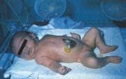
🔍 Figure: Exomphalos (omphalocele) – a midline abdominal wall defect with a membranous sac (amnion and peritoneum) covering the herniated abdominal contents. The umbilical cord inserts into the sac. Contents include small bowel and liver. This is an exomphalos major.
💡 Examiner instruction (interactive): The candidate will be asked to identify exomphalos, differentiate from gastroschisis, discuss embryology (failure of lateral body wall folding), list associated anomalies (Beckwith-Wiedemann syndrome, cardiac defects, chromosomal anomalies), outline immediate management (bowel bag, NG tube, antibiotics, fluid resuscitation), and discuss surgical closure (primary vs staged).
🔍 Examiner Questions (interactive) – Click to reveal model answers
❓ Q1 (Examiner): “Describe the findings in the image. What is the most likely diagnosis? Differentiate from gastroschisis.”
✅ Candidate's structured answer:
• Findings: Midline, central abdominal wall defect, with a membranous sac (amnion and peritoneum) covering the herniated viscera (bowel and liver). Umbilical cord inserts into the sac.
• Diagnosis: Exomphalos (omphalocele).
• Difference from gastroschisis:
- Omphalocele: Midline defect, sac covers viscera, cord inserts into sac, associated anomalies common (cardiac, chromosomal, Beckwith-Wiedemann), may contain liver.
- Gastroschisis: Right-sided defect, no sac, bowel is exposed, cord normal insertion, rarely associated anomalies.
• Findings: Midline, central abdominal wall defect, with a membranous sac (amnion and peritoneum) covering the herniated viscera (bowel and liver). Umbilical cord inserts into the sac.
• Diagnosis: Exomphalos (omphalocele).
• Difference from gastroschisis:
- Omphalocele: Midline defect, sac covers viscera, cord inserts into sac, associated anomalies common (cardiac, chromosomal, Beckwith-Wiedemann), may contain liver.
- Gastroschisis: Right-sided defect, no sac, bowel is exposed, cord normal insertion, rarely associated anomalies.
❓ Q2 (Examiner): “What is the embryological basis of omphalocele?”
✅ Candidate's answer:
• Failure of the lateral body wall folds to close during the 4th-5th week of gestation → herniation of abdominal contents through the umbilical ring.
• The midgut normally herniates into the umbilical cord (physiological herniation) at 6-8 weeks and returns by 10-12 weeks. In omphalocele, the intestines (and sometimes liver) fail to return to the abdominal cavity.
• The herniated viscera are covered by a sac composed of amnion and peritoneum.
• The umbilical cord inserts into the apex of the sac.
• Failure of the lateral body wall folds to close during the 4th-5th week of gestation → herniation of abdominal contents through the umbilical ring.
• The midgut normally herniates into the umbilical cord (physiological herniation) at 6-8 weeks and returns by 10-12 weeks. In omphalocele, the intestines (and sometimes liver) fail to return to the abdominal cavity.
• The herniated viscera are covered by a sac composed of amnion and peritoneum.
• The umbilical cord inserts into the apex of the sac.
❓ Q3 (Examiner): “How do you classify omphalocele? What is the difference between exomphalos major and minor?”
✅ Candidate's answer:
• Exomphalos minor (small omphalocele): Defect <5 cm, contains only small bowel, usually can undergo primary fascial closure.
• Exomphalos major (giant omphalocele): Defect >5 cm, contains liver plus other viscera, abdominal cavity is small, primary closure is not possible (viscero-abdominal disproportion). Requires staged closure (silo) or conservative management.
• This infant has exomphalos major (liver-containing, large sac).
• Exomphalos minor (small omphalocele): Defect <5 cm, contains only small bowel, usually can undergo primary fascial closure.
• Exomphalos major (giant omphalocele): Defect >5 cm, contains liver plus other viscera, abdominal cavity is small, primary closure is not possible (viscero-abdominal disproportion). Requires staged closure (silo) or conservative management.
• This infant has exomphalos major (liver-containing, large sac).
❓ Q4 (Examiner): “What are the common associated anomalies with omphalocele? What syndromes are associated?”
✅ Candidate's answer:
• Associated anomalies occur in 50-70% of omphalocele cases (much higher than gastroschisis).
• Cardiac defects (most common, 30-50%): VSD, ASD, tetralogy of Fallot, hypoplastic left heart syndrome.
• Chromosomal anomalies (30%): Trisomy 13, 18, 21.
• Beckwith-Wiedemann syndrome (10-15%): Visceromegaly, macroglossia, macrosomia, hypoglycemia, ear creases/pits, increased risk of Wilms tumor, hepatoblastoma.
• Pentalogy of Cantrell: Omphalocele + sternal cleft + diaphragmatic hernia + pericardial defect + intracardiac anomaly.
• Cloacal exstrophy, OEIS complex (Omphalocele, Exstrophy, Imperforate anus, Spinal defects).
• Genetic syndromes: Beckwith-Wiedemann (11p15.5 imprinting), Trisomies.
• Associated anomalies occur in 50-70% of omphalocele cases (much higher than gastroschisis).
• Cardiac defects (most common, 30-50%): VSD, ASD, tetralogy of Fallot, hypoplastic left heart syndrome.
• Chromosomal anomalies (30%): Trisomy 13, 18, 21.
• Beckwith-Wiedemann syndrome (10-15%): Visceromegaly, macroglossia, macrosomia, hypoglycemia, ear creases/pits, increased risk of Wilms tumor, hepatoblastoma.
• Pentalogy of Cantrell: Omphalocele + sternal cleft + diaphragmatic hernia + pericardial defect + intracardiac anomaly.
• Cloacal exstrophy, OEIS complex (Omphalocele, Exstrophy, Imperforate anus, Spinal defects).
• Genetic syndromes: Beckwith-Wiedemann (11p15.5 imprinting), Trisomies.
❓ Q5 (Examiner): “What is your immediate management of this newborn in the delivery room and NICU?”
✅ Candidate's structured answer:
1️⃣ Protect the sac: Cover with sterile, moist, non-adherent dressing (saline-soaked gauze) and then a sterile plastic bag (bowel bag) to prevent drying, rupture, and infection. Do NOT attempt to reduce the sac.
2️⃣ Place infant in a sterile plastic bag (from neck down) to maintain warmth.
3️⃣ Orogastric (OG) tube placement to decompress the stomach, prevent aspiration, and reduce distention, on continuous low suction.
4️⃣ IV fluids and broad-spectrum IV antibiotics (ampicillin + gentamicin + metronidazole).
5️⃣ Monitoring: Blood glucose (hypoglycemia – suspect Beckwith-Wiedemann), temperature, blood pressure.
6️⃣ Emergency investigations: Blood gas, electrolytes, blood glucose. Echocardiogram, renal ultrasound, skeletal survey (for associated anomalies).
7️⃣ Urgent pediatric surgery consultation.
1️⃣ Protect the sac: Cover with sterile, moist, non-adherent dressing (saline-soaked gauze) and then a sterile plastic bag (bowel bag) to prevent drying, rupture, and infection. Do NOT attempt to reduce the sac.
2️⃣ Place infant in a sterile plastic bag (from neck down) to maintain warmth.
3️⃣ Orogastric (OG) tube placement to decompress the stomach, prevent aspiration, and reduce distention, on continuous low suction.
4️⃣ IV fluids and broad-spectrum IV antibiotics (ampicillin + gentamicin + metronidazole).
5️⃣ Monitoring: Blood glucose (hypoglycemia – suspect Beckwith-Wiedemann), temperature, blood pressure.
6️⃣ Emergency investigations: Blood gas, electrolytes, blood glucose. Echocardiogram, renal ultrasound, skeletal survey (for associated anomalies).
7️⃣ Urgent pediatric surgery consultation.
❓ Q6 (Examiner): “Why is it important to monitor glucose in an infant with omphalocele? What is the management?”
✅ Candidate's answer:
• Hypoglycemia risk: Due to Beckwith-Wiedemann syndrome (associated with omphalocele). BWS causes neonatal hyperinsulinism → severe, refractory hypoglycemia.
• Management:
- Frequent blood glucose monitoring (every 1-2 hours initially).
- IV dextrose infusion (starting at 6-8 mg/kg/min) if hypoglycemic.
- Diazoxide (10-15 mg/kg/day) for hyperinsulinism.
- If refractory, consider glucagon or octreotide.
- Untreated hypoglycemia causes brain injury.
• Hypoglycemia risk: Due to Beckwith-Wiedemann syndrome (associated with omphalocele). BWS causes neonatal hyperinsulinism → severe, refractory hypoglycemia.
• Management:
- Frequent blood glucose monitoring (every 1-2 hours initially).
- IV dextrose infusion (starting at 6-8 mg/kg/min) if hypoglycemic.
- Diazoxide (10-15 mg/kg/day) for hyperinsulinism.
- If refractory, consider glucagon or octreotide.
- Untreated hypoglycemia causes brain injury.
❓ Q7 (Examiner): “What are the surgical options for omphalocele closure? When is primary closure possible?”
✅ Candidate's answer:
• Primary fascial closure: Possible for small (<5 cm) defects (exomphalos minor) after reduction of viscera. Performed in the operating room.
• Staged closure (silo placement): For exomphalos major (large defect, liver-containing) due to viscero-abdominal disproportion. A spring-loaded or preformed silo is placed over the sac to gradually reduce the contents over 3-10 days by gravity and gentle compression, followed by delayed primary closure.
• Conservative (non-surgical) management: For giant omphalocele or infants with severe associated anomalies/inoperable cardiac disease. The sac is left intact, covered with antiseptic (silver sulfadiazine or betadine) to allow epithelialization. Closure is delayed for months (secondary intention), then later repair of hernia.
• Prosthetic mesh (Gore-Tex) may be used for large fascial defects.
• Primary fascial closure: Possible for small (<5 cm) defects (exomphalos minor) after reduction of viscera. Performed in the operating room.
• Staged closure (silo placement): For exomphalos major (large defect, liver-containing) due to viscero-abdominal disproportion. A spring-loaded or preformed silo is placed over the sac to gradually reduce the contents over 3-10 days by gravity and gentle compression, followed by delayed primary closure.
• Conservative (non-surgical) management: For giant omphalocele or infants with severe associated anomalies/inoperable cardiac disease. The sac is left intact, covered with antiseptic (silver sulfadiazine or betadine) to allow epithelialization. Closure is delayed for months (secondary intention), then later repair of hernia.
• Prosthetic mesh (Gore-Tex) may be used for large fascial defects.
❓ Q8 (Examiner): “What is Beckwith-Wiedemann syndrome? Why is it associated with omphalocele? What other features are seen?”
✅ Candidate's answer:
• Beckwith-Wiedemann syndrome (BWS): Overgrowth syndrome due to abnormal imprinting on chromosome 11p15.5 (IGF2 and CDKN1C genes).
• Features:
- Macroglossia (large tongue) – as seen in this infant.
- Omphalocele or umbilical hernia (50% of BWS).
- Macrosomia (birth weight >90th percentile).
- Neonatal hypoglycemia (hyperinsulinism).
- Ear creases/pits, nevus flammeus (facial).
- Visceromegaly (hepatomegaly, nephromegaly).
- Hemihyperplasia (asymmetric limb overgrowth).
- Tumor risk: Wilms tumor (nephroblastoma), hepatoblastoma, neuroblastoma, adrenocortical carcinoma. Surveillance: abdominal ultrasound every 3 months until age 7 years, serum AFP every 3 months until age 4 years.
• Omphalocele in BWS results from the same embryopathy (midline defect).
• Beckwith-Wiedemann syndrome (BWS): Overgrowth syndrome due to abnormal imprinting on chromosome 11p15.5 (IGF2 and CDKN1C genes).
• Features:
- Macroglossia (large tongue) – as seen in this infant.
- Omphalocele or umbilical hernia (50% of BWS).
- Macrosomia (birth weight >90th percentile).
- Neonatal hypoglycemia (hyperinsulinism).
- Ear creases/pits, nevus flammeus (facial).
- Visceromegaly (hepatomegaly, nephromegaly).
- Hemihyperplasia (asymmetric limb overgrowth).
- Tumor risk: Wilms tumor (nephroblastoma), hepatoblastoma, neuroblastoma, adrenocortical carcinoma. Surveillance: abdominal ultrasound every 3 months until age 7 years, serum AFP every 3 months until age 4 years.
• Omphalocele in BWS results from the same embryopathy (midline defect).
❓ Q9 (Examiner): “What investigations should be performed to screen for associated anomalies in a newborn with omphalocele?”
✅ Candidate's answer:
1️⃣ Echocardiogram – to rule out congenital heart disease (30-50%).
2️⃣ Renal ultrasound – to detect renal anomalies (agenesis, cystic dysplasia).
3️⃣ Spine X-ray or ultrasound – for vertebral anomalies, neural tube defects.
4️⃣ Karyotype / chromosomal microarray – for trisomies 13, 18, 21 (especially if other dysmorphic features).
5️⃣ Methylation analysis (11p15.5) – for Beckwith-Wiedemann syndrome (if macroglossia, macrosomia, hypoglycemia, or ear creases present).
6️⃣ Ophthalmology exam – if BWS suspected (routine).
7️⃣ Limb and facial examination – for hemihyperplasia, nevus flammeus.
1️⃣ Echocardiogram – to rule out congenital heart disease (30-50%).
2️⃣ Renal ultrasound – to detect renal anomalies (agenesis, cystic dysplasia).
3️⃣ Spine X-ray or ultrasound – for vertebral anomalies, neural tube defects.
4️⃣ Karyotype / chromosomal microarray – for trisomies 13, 18, 21 (especially if other dysmorphic features).
5️⃣ Methylation analysis (11p15.5) – for Beckwith-Wiedemann syndrome (if macroglossia, macrosomia, hypoglycemia, or ear creases present).
6️⃣ Ophthalmology exam – if BWS suspected (routine).
7️⃣ Limb and facial examination – for hemihyperplasia, nevus flammeus.
❓ Q10 (Examiner): “How do you manage feeding in an infant with omphalocele? When can enteral feeding start?”
✅ Candidate's answer:
• Initially NPO (nil per oral) – NG/OG tube on suction to decompress stomach. Start IV fluids and parenteral nutrition.
• After surgical closure: Ileus is common; enteral feeds are started when bowel function returns (passage of flatus/stool, decreasing NG output). Typically 5-10 days after primary closure, longer after silo/staged closure.
• Breast milk or formula – start with small volume continuous feeds, then progress to bolus.
• Slow advancement: May have feeding intolerance, GERD, delayed gastric emptying.
• If gastrostomy tube placed at time of repair, use for enteral feeding.
• In BWS with macroglossia: Feeding difficulty due to large tongue may require nasogastric or gastrostomy feeds temporarily.
• Initially NPO (nil per oral) – NG/OG tube on suction to decompress stomach. Start IV fluids and parenteral nutrition.
• After surgical closure: Ileus is common; enteral feeds are started when bowel function returns (passage of flatus/stool, decreasing NG output). Typically 5-10 days after primary closure, longer after silo/staged closure.
• Breast milk or formula – start with small volume continuous feeds, then progress to bolus.
• Slow advancement: May have feeding intolerance, GERD, delayed gastric emptying.
• If gastrostomy tube placed at time of repair, use for enteral feeding.
• In BWS with macroglossia: Feeding difficulty due to large tongue may require nasogastric or gastrostomy feeds temporarily.
❓ Q11 (Examiner): “What is the long-term prognosis for an infant with isolated exomphalos minor vs exomphalos major with Beckwith-Wiedemann syndrome?”
✅ Candidate's answer:
• Isolated exomphalos minor (small defect, no other anomalies): Excellent prognosis. Normal survival, normal gastrointestinal function, good cosmetic outcome after primary closure.
• Exomphalos major (giant, liver-containing): Higher morbidity (prolonged ventilation, feeding difficulties, abdominal compartment syndrome). Overall survival >90% with modern care, but may have longer hospital stay and need for silo/staged closure.
• Beckwith-Wiedemann syndrome: Prognosis depends on tumor surveillance. Normal intelligence (85-90%). Tumor risk (Wilms, hepatoblastoma) requires strict surveillance (abdominal US q3 months, AFP q3 months). Hypoglycemia usually resolves by infancy.
• Associated cardiac or chromosomal anomalies: Prognosis determined by those anomalies.
• Long-term follow-up: Multidisciplinary (pediatric surgery, genetics, oncology (tumor surveillance), endocrinology (hypoglycemia), cardiology, developmental pediatrics).
• Isolated exomphalos minor (small defect, no other anomalies): Excellent prognosis. Normal survival, normal gastrointestinal function, good cosmetic outcome after primary closure.
• Exomphalos major (giant, liver-containing): Higher morbidity (prolonged ventilation, feeding difficulties, abdominal compartment syndrome). Overall survival >90% with modern care, but may have longer hospital stay and need for silo/staged closure.
• Beckwith-Wiedemann syndrome: Prognosis depends on tumor surveillance. Normal intelligence (85-90%). Tumor risk (Wilms, hepatoblastoma) requires strict surveillance (abdominal US q3 months, AFP q3 months). Hypoglycemia usually resolves by infancy.
• Associated cardiac or chromosomal anomalies: Prognosis determined by those anomalies.
• Long-term follow-up: Multidisciplinary (pediatric surgery, genetics, oncology (tumor surveillance), endocrinology (hypoglycemia), cardiology, developmental pediatrics).
❓ Q12 (Examiner): “Can omphalocele be diagnosed prenatally? What are the ultrasound findings?”
✅ Candidate's answer:
• Yes, commonly diagnosed prenatally at 18-22 weeks ultrasound.
• Findings:
- Midline defect with herniated viscera (liver, bowel) covered by a membrane.
- Umbilical cord insertion into the sac.
- Elevated maternal serum AFP (but not diagnostic).
- Associated anomalies: cardiac (most common), neural tube defects, chromosomal abnormalities.
- Signs of Beckwith-Wiedemann: Polyhydramnios, macrosomia, placentomegaly, macroglossia, visceromegaly.
• Management: Detailed anomaly scan, fetal echocardiogram, amniocentesis for karyotype/microarray (trisomy 13,18,21), methylation analysis for BWS if features present.
• Counsel parents: Plan delivery at tertiary center with NICU, pediatric surgery, and subspecialties.
• Yes, commonly diagnosed prenatally at 18-22 weeks ultrasound.
• Findings:
- Midline defect with herniated viscera (liver, bowel) covered by a membrane.
- Umbilical cord insertion into the sac.
- Elevated maternal serum AFP (but not diagnostic).
- Associated anomalies: cardiac (most common), neural tube defects, chromosomal abnormalities.
- Signs of Beckwith-Wiedemann: Polyhydramnios, macrosomia, placentomegaly, macroglossia, visceromegaly.
• Management: Detailed anomaly scan, fetal echocardiogram, amniocentesis for karyotype/microarray (trisomy 13,18,21), methylation analysis for BWS if features present.
• Counsel parents: Plan delivery at tertiary center with NICU, pediatric surgery, and subspecialties.
❓ Q13 (Examiner): “What are the complications of omphalocele repair?”
✅ Candidate's answer:
• Early complications:
- Abdominal compartment syndrome (tight closure → increased intra-abdominal pressure → renal failure, respiratory compromise, bowel ischemia).
- Wound infection, dehiscence, sac rupture (if not protected).
- Sepsis, prolonged ileus, necrotizing enterocolitis.
- Respiratory failure (due to decreased diaphragmatic excursion).
• Late complications:
- Recurrent omphalocele / incisional hernia – may require repeat repair.
- Intestinal obstruction (adhesions, malrotation).
- GERD and feeding intolerance.
- Cosmetic deformity (scar, lack of umbilicus).
• In BWS: Late-onset tumors (Wilms, hepatoblastoma) – requires surveillance.
• Early complications:
- Abdominal compartment syndrome (tight closure → increased intra-abdominal pressure → renal failure, respiratory compromise, bowel ischemia).
- Wound infection, dehiscence, sac rupture (if not protected).
- Sepsis, prolonged ileus, necrotizing enterocolitis.
- Respiratory failure (due to decreased diaphragmatic excursion).
• Late complications:
- Recurrent omphalocele / incisional hernia – may require repeat repair.
- Intestinal obstruction (adhesions, malrotation).
- GERD and feeding intolerance.
- Cosmetic deformity (scar, lack of umbilicus).
• In BWS: Late-onset tumors (Wilms, hepatoblastoma) – requires surveillance.
❓ Q14 (Examiner): “How will you counsel the parents of this newborn with exomphalos major and macroglossia (suspected Beckwith-Wiedemann syndrome)?”
✅ Candidate's structured answer:
• “Your baby has a condition called exomphalos (omphalocele) – a hole in the middle of the belly where the intestines and liver are covered by a sac. It is a rare birth defect.”
• “The large tongue (macroglossia) and large size suggest an associated condition called Beckwith-Wiedemann syndrome – an overgrowth syndrome that increases the risk of low blood sugar (hypoglycemia) and later kidney or liver tumors.”
• “We will protect the sac, start IV fluids, antibiotics, and monitor blood sugar closely.”
• “Surgery to put the bowel back into the belly may be done in one operation or gradually over several days using a plastic silo (a temporary pouch). We will work with the pediatric surgeon to decide the best approach.”
• “The outlook is good. Most babies survive, especially if there are no major heart defects. Your baby will need long-term follow-up: blood sugar management, tumor screening (ultrasounds every 3 months until age 7 years), and checkups for growth and development.”
• “You did nothing wrong – this is not anyone’s fault. We have a team of specialists (neonatology, surgery, genetics, endocrinology, oncology) to support your baby and your family.”
• “Your baby has a condition called exomphalos (omphalocele) – a hole in the middle of the belly where the intestines and liver are covered by a sac. It is a rare birth defect.”
• “The large tongue (macroglossia) and large size suggest an associated condition called Beckwith-Wiedemann syndrome – an overgrowth syndrome that increases the risk of low blood sugar (hypoglycemia) and later kidney or liver tumors.”
• “We will protect the sac, start IV fluids, antibiotics, and monitor blood sugar closely.”
• “Surgery to put the bowel back into the belly may be done in one operation or gradually over several days using a plastic silo (a temporary pouch). We will work with the pediatric surgeon to decide the best approach.”
• “The outlook is good. Most babies survive, especially if there are no major heart defects. Your baby will need long-term follow-up: blood sugar management, tumor screening (ultrasounds every 3 months until age 7 years), and checkups for growth and development.”
• “You did nothing wrong – this is not anyone’s fault. We have a team of specialists (neonatology, surgery, genetics, endocrinology, oncology) to support your baby and your family.”
🗣️ Examiner's probing / high-yield points:
• "What is the key difference between omphalocele and gastroschisis?" → Omphalocele has a sac, cord inserts into sac, midline; gastroschisis: no sac, right-sided, normal cord insertion.
• "What is the most common associated anomaly?" → Cardiac defects (30-50%).
• "What syndrome is suggested by macroglossia + omphalocele + macrosomia?" → Beckwith-Wiedemann syndrome.
• "Why is hypoglycemia a concern?" → Hyperinsulinism in BWS → treat with glucose and diazoxide.
• "What tumor surveillance is needed in BWS?" → Abdominal US q3 months until age 7 years; AFP q3 months until age 4 years.
• "What is exomphalos major?" → Defect >5 cm, contains liver, requires staged closure.
• "What is pentalogy of Cantrell?" → Omphalocele + sternal cleft + diaphragmatic hernia + pericardial defect + cardiac anomaly.
• "What is the key difference between omphalocele and gastroschisis?" → Omphalocele has a sac, cord inserts into sac, midline; gastroschisis: no sac, right-sided, normal cord insertion.
• "What is the most common associated anomaly?" → Cardiac defects (30-50%).
• "What syndrome is suggested by macroglossia + omphalocele + macrosomia?" → Beckwith-Wiedemann syndrome.
• "Why is hypoglycemia a concern?" → Hyperinsulinism in BWS → treat with glucose and diazoxide.
• "What tumor surveillance is needed in BWS?" → Abdominal US q3 months until age 7 years; AFP q3 months until age 4 years.
• "What is exomphalos major?" → Defect >5 cm, contains liver, requires staged closure.
• "What is pentalogy of Cantrell?" → Omphalocele + sternal cleft + diaphragmatic hernia + pericardial defect + cardiac anomaly.
📘 Exomphalos (Omphalocele) – Core Revision for TOACS
🔍 Definition
Midline abdominal wall defect covered by a membranous sac (amnion and peritoneum). Umbilical cord inserts into the sac. Incidence 1 in 4,000-5,000 live births.
Midline abdominal wall defect covered by a membranous sac (amnion and peritoneum). Umbilical cord inserts into the sac. Incidence 1 in 4,000-5,000 live births.
📊 Classification
Minor (<5 cm, bowel only) – primary closure. Major (>5 cm, contains liver) – staged closure (silo) or conservative management.
Minor (<5 cm, bowel only) – primary closure. Major (>5 cm, contains liver) – staged closure (silo) or conservative management.
🧬 Associated Anomalies
Cardiac (30-50%), chromosomal (trisomy 13,18,21), Beckwith-Wiedemann (11p15.5), Pentalogy of Cantrell, OEIS complex.
Cardiac (30-50%), chromosomal (trisomy 13,18,21), Beckwith-Wiedemann (11p15.5), Pentalogy of Cantrell, OEIS complex.
🩺 Immediate Management
Protect sac (moist + plastic bag), NG tube, IV fluids, antibiotics, glucose monitoring (hypoglycemia in BWS).
Protect sac (moist + plastic bag), NG tube, IV fluids, antibiotics, glucose monitoring (hypoglycemia in BWS).
⚕️ Surgical Repair
Primary closure (small defects). Staged silo closure (giant omphalocele). Conservative epithelialization for high-risk infants.
Primary closure (small defects). Staged silo closure (giant omphalocele). Conservative epithelialization for high-risk infants.
📈 Beckwith-Wiedemann Syndrome
Macroglossia, macrosomia, omphalocele, hypoglycemia, ear creases, tumor risk (Wilms, hepatoblastoma). Surveillance: abdominal US q3 months until age 7 years, AFP q3 months until age 4 years.
Macroglossia, macrosomia, omphalocele, hypoglycemia, ear creases, tumor risk (Wilms, hepatoblastoma). Surveillance: abdominal US q3 months until age 7 years, AFP q3 months until age 4 years.
⭐ High-yield pearls for TOACS (Exomphalos):
• Sac + midline + cord insertion = omphalocele. No sac + right-sided + normal cord = gastroschisis.
• Most common associated anomaly: Congenital heart disease (30-50%).
• Beckwith-Wiedemann syndrome – macroglossia, macrosomia, hypoglycemia, tumor risk.
• Do NOT rupture the sac. Cover with moist dressing + plastic bag.
• Monitor blood glucose – hypoglycemia indicates hyperinsulinism (BWS).
• Exomphalos major (giant) requires staged silo closure. Primary closure impossible due to viscero-abdominal disproportion.
• Tumor surveillance in BWS: Abdominal US q3 months until age 7; AFP q3 months until age 4.
• Sac + midline + cord insertion = omphalocele. No sac + right-sided + normal cord = gastroschisis.
• Most common associated anomaly: Congenital heart disease (30-50%).
• Beckwith-Wiedemann syndrome – macroglossia, macrosomia, hypoglycemia, tumor risk.
• Do NOT rupture the sac. Cover with moist dressing + plastic bag.
• Monitor blood glucose – hypoglycemia indicates hyperinsulinism (BWS).
• Exomphalos major (giant) requires staged silo closure. Primary closure impossible due to viscero-abdominal disproportion.
• Tumor surveillance in BWS: Abdominal US q3 months until age 7; AFP q3 months until age 4.
🗣️ Candidate's role-play & examiner feedback
💬 To the candidate (role‑play): You will be asked the 14 questions from the Examiner Q&A tab (including clinical recognition, differentiation from gastroschisis, embryology, associated anomalies, Beckwith-Wiedemann syndrome, immediate management, surgical options, and parental counseling). Provide concise, evidence‑based answers. Examiner may ask about the differences between exomphalos minor and major or the tumor surveillance in BWS. Use structured points and demonstrate empathy when counseling parents.
📝 Examiner Marking Grid (Exomphalos – TOACS station):
- ✅ Correctly identifies omphalocele from image (sac, midline, cord insertion)
- ✅ Differentiates from gastroschisis (sac vs no sac, midline vs right-sided)
- ✅ Explains embryology (failure of lateral body wall folding)
- ✅ Lists associated anomalies (cardiac, chromosomal, Beckwith-Wiedemann)
- ✅ Recognizes features of Beckwith-Wiedemann syndrome (macroglossia, macrosomia, hypoglycemia risk)
- ✅ Outlines immediate management (protect sac, NG tube, IV fluids, antibiotics)
- ✅ Discusses surgical options (primary vs staged silo closure)
- ✅ States that exomphalos major (liver-containing) requires staged closure
- ✅ Describes tumor surveillance in BWS (abdominal US q3 months, AFP q3 months)
- ✅ Provides appropriate parental counseling (reassurance, multidisciplinary follow-up)
📚 Key references: Nelson Textbook of Pediatrics 22e (Chapter 385.6 – Omphalocele), Beckwith-Wiedemann syndrome guidelines, CPSP guidelines on abdominal wall defects, Journal of Pediatric Surgery.