FCPS Paediatrics TOACS · Interactive Station
🩺 Hirschsprung Disease — Congenital aganglionic megacolon, failure to pass meconium (4-5 days), abdominal distension, enterocolitis, contrast enema, rectal biopsy, surgical management (Swenson, Soave), associated syndromes (Trisomy 21)
📚 Paeds Online – paeds.online
⚕️ OBSERVED STATION · CPSP FORMAT · 8 MINUTES · SEPARATE TABS · CLINICAL PHOTO INCLUDED
📖 Problem-oriented Clinical Scenario + Photograph
👶🏻 Clinical Scenario (read aloud – 2 min):
A 5-day-old term male infant is brought to the emergency department by his parents with concerns of "not having a proper poop since birth." The infant passed a small, pellet-like meconium on day 2 but has passed no further stool. Over the last 24 hours, he has developed progressive abdominal distension, bilious vomiting, and poor feeding. On examination, the infant is mildly dehydrated, abdomen is distended, tense, and tympanitic. There is no visible peristalsis. A gentle rectal examination is performed – the anal sphincter is tight, and after withdrawal of the finger, a large explosive passage of gas and meconium occurs (explosive stool sign). The infant becomes transiently more comfortable. The mother is anxious and asks, "Why hasn't my baby pooped normally? Is it serious? He is already 5 days old." A clinical photograph of a distended abdomen in a neonate with Hirschsprung disease is shown.
A 5-day-old term male infant is brought to the emergency department by his parents with concerns of "not having a proper poop since birth." The infant passed a small, pellet-like meconium on day 2 but has passed no further stool. Over the last 24 hours, he has developed progressive abdominal distension, bilious vomiting, and poor feeding. On examination, the infant is mildly dehydrated, abdomen is distended, tense, and tympanitic. There is no visible peristalsis. A gentle rectal examination is performed – the anal sphincter is tight, and after withdrawal of the finger, a large explosive passage of gas and meconium occurs (explosive stool sign). The infant becomes transiently more comfortable. The mother is anxious and asks, "Why hasn't my baby pooped normally? Is it serious? He is already 5 days old." A clinical photograph of a distended abdomen in a neonate with Hirschsprung disease is shown.
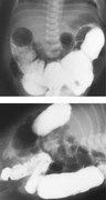
🔍 Figure: Abdominal distension in a 5-day-old neonate with Hirschsprung disease. The child has failed to pass meconium adequately since birth. A contrast enema typically shows a transition zone (narrow distal aganglionic segment with proximal dilation). The image may demonstrate the classic radiographic findings.
💡 Examiner instruction (interactive): The candidate will be asked to diagnose Hirschsprung disease, describe the typical presentation (delayed passage of meconium beyond 48 hours – here 5 days, abdominal distension, explosive stool after rectal exam), order confirmatory tests (contrast enema, rectal suction biopsy), discuss the pathophysiology (absence of ganglion cells in Auerbach and Meissner plexuses), and outline surgical management (pull-through procedure). Also, discuss associated conditions (Trisomy 21) and risk of enterocolitis.
🔍 Examiner Questions (interactive) – Click to reveal model answers
❓ Q1 (Examiner): “What is the most likely diagnosis in this 5-day-old infant who has not passed meconium adequately since birth and has a distended abdomen with explosive stool after rectal exam? List the key clinical features of Hirschsprung disease in the newborn.”
✅ Candidate's answer:
• Diagnosis: Hirschsprung disease (congenital aganglionic megacolon).
• Key neonatal features:
1️⃣ Failure to pass meconium within 48 hours of birth – this infant is now 5 days old with only a small pellet on day 2 (classic).
2️⃣ Progressive abdominal distension (days 2-5).
3️⃣ Bilious vomiting (suggests distal obstruction).
4️⃣ Explosive passage of gas and stool after rectal examination (pathognomonic sign).
5️⃣ Tight anal sphincter on digital examination.
6️⃣ Enterocolitis (fever, diarrhea, shock, abdominal distension) – a life-threatening complication.
• Diagnosis: Hirschsprung disease (congenital aganglionic megacolon).
• Key neonatal features:
1️⃣ Failure to pass meconium within 48 hours of birth – this infant is now 5 days old with only a small pellet on day 2 (classic).
2️⃣ Progressive abdominal distension (days 2-5).
3️⃣ Bilious vomiting (suggests distal obstruction).
4️⃣ Explosive passage of gas and stool after rectal examination (pathognomonic sign).
5️⃣ Tight anal sphincter on digital examination.
6️⃣ Enterocolitis (fever, diarrhea, shock, abdominal distension) – a life-threatening complication.
❓ Q2 (Examiner): “Explain the pathophysiology of Hirschsprung disease. Which part of the colon is usually affected?”
✅ Candidate's answer:
• Pathophysiology: Failure of migration of neural crest cells (enteric ganglion cells) to the distal bowel during weeks 5-12 of gestation → absence of both Meissner (submucosal) and Auerbach (myenteric) plexuses in the affected segment.
• Consequently, the aganglionic segment remains tonically contracted (spastic) → functional obstruction, proximal bowel dilates (megacolon).
• Most common (75-80%): Short-segment disease – aganglionosis confined to rectosigmoid colon.
• Long-segment disease (10-15%): extends proximal to sigmoid.
• Total colonic aganglionosis (5-10%): entire colon involved.
• Genetic basis: RET proto-oncogene mutations (>50% familial cases), also EDNRB, EDN3, SOX10.
• Pathophysiology: Failure of migration of neural crest cells (enteric ganglion cells) to the distal bowel during weeks 5-12 of gestation → absence of both Meissner (submucosal) and Auerbach (myenteric) plexuses in the affected segment.
• Consequently, the aganglionic segment remains tonically contracted (spastic) → functional obstruction, proximal bowel dilates (megacolon).
• Most common (75-80%): Short-segment disease – aganglionosis confined to rectosigmoid colon.
• Long-segment disease (10-15%): extends proximal to sigmoid.
• Total colonic aganglionosis (5-10%): entire colon involved.
• Genetic basis: RET proto-oncogene mutations (>50% familial cases), also EDNRB, EDN3, SOX10.
❓ Q3 (Examiner): “What is Hirschsprung-associated enterocolitis (HAEC)? How does it present and how is it treated?”
✅ Candidate's answer:
• HAEC: life-threatening inflammation of the bowel proximal to the aganglionic segment, often with mucosal ulceration, bacterial translocation, and sepsis.
• Presentation: explosive, foul-smelling watery diarrhea (may be mistaken for gastroenteritis), abdominal distension, fever, lethargy, hypotension, shock, septicemia.
• Treatment (medical emergency):
- Rectal irrigation with normal saline (decompression).
- Intravenous antibiotics (cover gram-negatives and anaerobes: metronidazole + gentamicin/cefotaxime).
- IV fluids, correction of electrolytes, and vasopressors if shock.
- Nasogastric decompression.
- Avoid barium enema during acute episode.
- Definitive surgery (pull-through) after stabilization.
• Recurrent enterocolitis may occur even after pull-through.
• HAEC: life-threatening inflammation of the bowel proximal to the aganglionic segment, often with mucosal ulceration, bacterial translocation, and sepsis.
• Presentation: explosive, foul-smelling watery diarrhea (may be mistaken for gastroenteritis), abdominal distension, fever, lethargy, hypotension, shock, septicemia.
• Treatment (medical emergency):
- Rectal irrigation with normal saline (decompression).
- Intravenous antibiotics (cover gram-negatives and anaerobes: metronidazole + gentamicin/cefotaxime).
- IV fluids, correction of electrolytes, and vasopressors if shock.
- Nasogastric decompression.
- Avoid barium enema during acute episode.
- Definitive surgery (pull-through) after stabilization.
• Recurrent enterocolitis may occur even after pull-through.
❓ Q4 (Examiner): “What investigations would you order to confirm Hirschsprung disease in this 5-day-old neonate?”
✅ Candidate's answer:
• 1. Abdominal X-ray (to rule out perforation, shows dilated bowel loops, no distal air).
• 2. Contrast enema (barium or water-soluble): Demonstrates a transition zone – narrow, irregular aganglionic distal segment with dilated proximal colon. Retained barium after 24 hours (delayed evacuation) supports diagnosis. Do not perform in acute enterocolitis (risk of perforation).
• 3. Rectal suction biopsy (gold standard): Performed at 2-3 cm above dentate line. Shows absence of ganglion cells and hypertrophic nerve trunks (acetylcholinesterase staining – positive thick nerve fibers in lamina propria and muscularis mucosae).
• 4. Anorectal manometry: Absence of rectoanal inhibitory reflex (RAIR) – reflex relaxation of internal anal sphincter with rectal distension is absent. Less reliable in neonates.
• 5. Full-thickness rectal biopsy (if suction biopsy inconclusive, or for definitive pathology prior to surgery).
• 1. Abdominal X-ray (to rule out perforation, shows dilated bowel loops, no distal air).
• 2. Contrast enema (barium or water-soluble): Demonstrates a transition zone – narrow, irregular aganglionic distal segment with dilated proximal colon. Retained barium after 24 hours (delayed evacuation) supports diagnosis. Do not perform in acute enterocolitis (risk of perforation).
• 3. Rectal suction biopsy (gold standard): Performed at 2-3 cm above dentate line. Shows absence of ganglion cells and hypertrophic nerve trunks (acetylcholinesterase staining – positive thick nerve fibers in lamina propria and muscularis mucosae).
• 4. Anorectal manometry: Absence of rectoanal inhibitory reflex (RAIR) – reflex relaxation of internal anal sphincter with rectal distension is absent. Less reliable in neonates.
• 5. Full-thickness rectal biopsy (if suction biopsy inconclusive, or for definitive pathology prior to surgery).
❓ Q5 (Examiner): “Describe the contrast enema findings in Hirschsprung disease. What is the transition zone?”
✅ Candidate's answer:
• Transition zone: A cone-shaped or funnel-shaped area where the narrow, spastic distal (aganglionic) segment meets the dilated proximal (ganglionic) colon.
• In short-segment disease, the transition zone is usually in the rectosigmoid.
• In long-segment disease, the transition zone is more proximal (splenic flexure, transverse colon).
• Other findings: Irregular contractions of aganglionic segment (sawtooth appearance), delayed evacuation of contrast (>24 hours).
• Important: In neonates, the transition zone may not be well-developed initially because the proximal bowel has not yet dilated significantly. Repeat enema after a few weeks may be helpful.
• Total colonic aganglionosis: Microcolon, no clear transition zone, shortened colon.
• Transition zone: A cone-shaped or funnel-shaped area where the narrow, spastic distal (aganglionic) segment meets the dilated proximal (ganglionic) colon.
• In short-segment disease, the transition zone is usually in the rectosigmoid.
• In long-segment disease, the transition zone is more proximal (splenic flexure, transverse colon).
• Other findings: Irregular contractions of aganglionic segment (sawtooth appearance), delayed evacuation of contrast (>24 hours).
• Important: In neonates, the transition zone may not be well-developed initially because the proximal bowel has not yet dilated significantly. Repeat enema after a few weeks may be helpful.
• Total colonic aganglionosis: Microcolon, no clear transition zone, shortened colon.
❓ Q6 (Examiner): “What do you expect to see on rectal suction biopsy in Hirschsprung disease? Which staining technique is used?”
✅ Candidate's answer:
• Findings:
- Absence of ganglion cells in submucosal (Meissner) and myenteric (Auerbach) plexuses.
- Presence of hypertrophic, non-myelinated nerve trunks in the lamina propria and muscularis mucosae.
• Staining: Acetylcholinesterase (AChE) histochemistry is the gold standard – shows strongly positive thick nerve fibers in the lamina propria and muscularis mucosae (normally absent or fine).
• Calretinin immunostaining: Absence of calretinin-positive ganglion cells and nerve fibers (rapid, reliable alternative).
• H&E staining: can identify ganglion cells but experienced pathologist required.
• Biopsy must be adequate (at least 3-5 mm of submucosa, 2-3 cm above dentate line).
• Findings:
- Absence of ganglion cells in submucosal (Meissner) and myenteric (Auerbach) plexuses.
- Presence of hypertrophic, non-myelinated nerve trunks in the lamina propria and muscularis mucosae.
• Staining: Acetylcholinesterase (AChE) histochemistry is the gold standard – shows strongly positive thick nerve fibers in the lamina propria and muscularis mucosae (normally absent or fine).
• Calretinin immunostaining: Absence of calretinin-positive ganglion cells and nerve fibers (rapid, reliable alternative).
• H&E staining: can identify ganglion cells but experienced pathologist required.
• Biopsy must be adequate (at least 3-5 mm of submucosa, 2-3 cm above dentate line).
❓ Q7 (Examiner): “What are the differential diagnoses for a neonate with delayed passage of meconium (5 days) and abdominal distension?”
✅ Candidate's answer:
• Meconium plug syndrome – contrast enema reveals meconium plugs, but ganglion cells present.
• Meconium ileus (cystic fibrosis) – distal small bowel obstruction, ground-glass appearance on X-ray, no transition zone.
• Small left colon syndrome – infants of diabetic mothers, transition at splenic flexure; usually resolves spontaneously.
• Intestinal atresia / stenosis – X-ray shows complete obstruction, no contrast passage.
• Necrotizing enterocolitis (NEC) – pneumatosis, clinical sepsis, usually later onset (but can occur in first week).
• Hypothyroidism – constipation, but not explosive stool after rectal exam.
• Functional constipation (rare in first 48h, but possible by day 5).
• Rectal biopsy differentiates.
• Meconium plug syndrome – contrast enema reveals meconium plugs, but ganglion cells present.
• Meconium ileus (cystic fibrosis) – distal small bowel obstruction, ground-glass appearance on X-ray, no transition zone.
• Small left colon syndrome – infants of diabetic mothers, transition at splenic flexure; usually resolves spontaneously.
• Intestinal atresia / stenosis – X-ray shows complete obstruction, no contrast passage.
• Necrotizing enterocolitis (NEC) – pneumatosis, clinical sepsis, usually later onset (but can occur in first week).
• Hypothyroidism – constipation, but not explosive stool after rectal exam.
• Functional constipation (rare in first 48h, but possible by day 5).
• Rectal biopsy differentiates.
❓ Q8 (Examiner): “Which genetic syndromes are associated with Hirschsprung disease?”
✅ Candidate's answer:
• Down syndrome (Trisomy 21): Most common association – 2-10% of Down syndrome patients have Hirschsprung.
• Waardenburg syndrome (pigmentary anomalies, deafness) – due to EDNRB, EDN3, SOX10 mutations.
• MEN2A (multiple endocrine neoplasia type 2A): RET mutation – Hirschsprung + medullary thyroid carcinoma + pheochromocytoma.
• MEN2B: Hirschsprung + mucosal neuromas, marfanoid habitus.
• Bardet-Biedl syndrome (obesity, polydactyly, retinitis pigmentosa).
• Congenital central hypoventilation syndrome (CCHS, Ondine’s curse) – associated with RET, PHOX2B mutations.
• Goldberg-Shprintzen syndrome (microcephaly, intellectual disability).
• Mowat-Wilson syndrome.
• Down syndrome (Trisomy 21): Most common association – 2-10% of Down syndrome patients have Hirschsprung.
• Waardenburg syndrome (pigmentary anomalies, deafness) – due to EDNRB, EDN3, SOX10 mutations.
• MEN2A (multiple endocrine neoplasia type 2A): RET mutation – Hirschsprung + medullary thyroid carcinoma + pheochromocytoma.
• MEN2B: Hirschsprung + mucosal neuromas, marfanoid habitus.
• Bardet-Biedl syndrome (obesity, polydactyly, retinitis pigmentosa).
• Congenital central hypoventilation syndrome (CCHS, Ondine’s curse) – associated with RET, PHOX2B mutations.
• Goldberg-Shprintzen syndrome (microcephaly, intellectual disability).
• Mowat-Wilson syndrome.
❓ Q9 (Examiner): “What is the definitive treatment for Hirschsprung disease? Describe the timing and types of pull-through procedures.”
✅ Candidate's answer:
• Definitive treatment: Surgical resection of the aganglionic bowel and pull-through of normally ganglionic colon to the anus.
• Timing: Traditionally after 4-6 weeks of age (weight >4 kg). Now primary pull-through in the neonatal period (1-3 months) is increasingly performed if the infant is stable and no enterocolitis. Previously, a two-stage approach: initial leveling colostomy (in sick or very young infants) followed by pull-through at 6-12 months.
• Common pull-through procedures:
1️⃣ Swenson procedure: Resection of aganglionic segment + end-to-end anastomosis (preserves anal sphincter).
2️⃣ Soave procedure: Endorectal pull-through – aganglionic mucosa/submucosa resected, ganglionic bowel pulled through muscular cuff.
3️⃣ Duhamel procedure: Retrorectal pull-through with side-to-side anastomosis.
• Transanal pull-through (TEPT) is now preferred for short-segment disease (no abdominal incision).
• Laparoscopic-assisted pull-through for long-segment or total colonic.
• Post-operative complications: anastomotic leak, stricture, persistent constipation (residual aganglionosis), enterocolitis, soiling (incontinence).
• Definitive treatment: Surgical resection of the aganglionic bowel and pull-through of normally ganglionic colon to the anus.
• Timing: Traditionally after 4-6 weeks of age (weight >4 kg). Now primary pull-through in the neonatal period (1-3 months) is increasingly performed if the infant is stable and no enterocolitis. Previously, a two-stage approach: initial leveling colostomy (in sick or very young infants) followed by pull-through at 6-12 months.
• Common pull-through procedures:
1️⃣ Swenson procedure: Resection of aganglionic segment + end-to-end anastomosis (preserves anal sphincter).
2️⃣ Soave procedure: Endorectal pull-through – aganglionic mucosa/submucosa resected, ganglionic bowel pulled through muscular cuff.
3️⃣ Duhamel procedure: Retrorectal pull-through with side-to-side anastomosis.
• Transanal pull-through (TEPT) is now preferred for short-segment disease (no abdominal incision).
• Laparoscopic-assisted pull-through for long-segment or total colonic.
• Post-operative complications: anastomotic leak, stricture, persistent constipation (residual aganglionosis), enterocolitis, soiling (incontinence).
❓ Q10 (Examiner): “How does total colonic aganglionosis (TCA) differ from classic Hirschsprung? How is it diagnosed and managed?”
✅ Candidate's answer:
• TCA: Entire colon (and sometimes terminal ileum) is aganglionic; accounts for 5-10% of Hirschsprung.
• Presentation: Delayed meconium passage, severe abdominal distension, vomiting, and early enterocolitis.
• Diagnosis: Contrast enema shows a microcolon or “question mark” colon (short, straight, no transition zone). Rectal suction biopsy confirms absence of ganglion cells. Intraoperative biopsies of colon and ileum are required to determine level of aganglionosis.
• Management:
- Initial diversion (ileostomy) at the level of ganglionic bowel (usually terminal ileum).
- Pull-through procedure: Ileal (or right colon) pull-through with creation of a reservoir (cecal patch, J-pouch) to reduce diarrhea and malabsorption.
- Lifelong risk of: bacterial overgrowth, chronic diarrhea, failure to thrive, need for parenteral nutrition in some cases.
• TCA: Entire colon (and sometimes terminal ileum) is aganglionic; accounts for 5-10% of Hirschsprung.
• Presentation: Delayed meconium passage, severe abdominal distension, vomiting, and early enterocolitis.
• Diagnosis: Contrast enema shows a microcolon or “question mark” colon (short, straight, no transition zone). Rectal suction biopsy confirms absence of ganglion cells. Intraoperative biopsies of colon and ileum are required to determine level of aganglionosis.
• Management:
- Initial diversion (ileostomy) at the level of ganglionic bowel (usually terminal ileum).
- Pull-through procedure: Ileal (or right colon) pull-through with creation of a reservoir (cecal patch, J-pouch) to reduce diarrhea and malabsorption.
- Lifelong risk of: bacterial overgrowth, chronic diarrhea, failure to thrive, need for parenteral nutrition in some cases.
❓ Q11 (Examiner): “What are the long-term complications after pull-through surgery for Hirschsprung disease?”
✅ Candidate's answer:
• Enterocolitis (post-pull-through) – can occur in 20-30% even after successful surgery; may require rectal irrigations, antibiotics.
• Persistent obstructive symptoms – residual aganglionosis (if inadequate resection), achalasia of the internal anal sphincter, or anastomotic stricture.
• Constipation and soiling (fecal incontinence) – common, especially in long-segment disease; requires bowel management program.
• Urinary dysfunction (due to pelvic nerve injury during surgery).
• Growth failure, malnutrition (especially total colonic aganglionosis).
• Psychological issues (social stigma, toilet training difficulties).
• Risk of medullary thyroid carcinoma in MEN2A associated cases – requires prophylactic thyroidectomy in childhood.
• Enterocolitis (post-pull-through) – can occur in 20-30% even after successful surgery; may require rectal irrigations, antibiotics.
• Persistent obstructive symptoms – residual aganglionosis (if inadequate resection), achalasia of the internal anal sphincter, or anastomotic stricture.
• Constipation and soiling (fecal incontinence) – common, especially in long-segment disease; requires bowel management program.
• Urinary dysfunction (due to pelvic nerve injury during surgery).
• Growth failure, malnutrition (especially total colonic aganglionosis).
• Psychological issues (social stigma, toilet training difficulties).
• Risk of medullary thyroid carcinoma in MEN2A associated cases – requires prophylactic thyroidectomy in childhood.
❓ Q12 (Examiner): “What is the role of anorectal manometry in diagnosing Hirschsprung disease? What finding is expected?”
✅ Candidate's answer:
• Anorectal manometry measures pressures in the anal canal and rectal response to distension.
• Normal response: Rectal distension → reflex relaxation of internal anal sphincter (rectoanal inhibitory reflex, RAIR).
• In Hirschsprung disease: Absence of RAIR due to lack of inhibitory neurons (nitrergic) in the aganglionic segment.
• Utility: High sensitivity and specificity, especially in older infants and children. In neonates, the reflex may be immature, and false positives can occur, so it is not the preferred first-line test in newborns.
• Advantages: Noninvasive, no radiation, can be done as outpatient.
• Anorectal manometry measures pressures in the anal canal and rectal response to distension.
• Normal response: Rectal distension → reflex relaxation of internal anal sphincter (rectoanal inhibitory reflex, RAIR).
• In Hirschsprung disease: Absence of RAIR due to lack of inhibitory neurons (nitrergic) in the aganglionic segment.
• Utility: High sensitivity and specificity, especially in older infants and children. In neonates, the reflex may be immature, and false positives can occur, so it is not the preferred first-line test in newborns.
• Advantages: Noninvasive, no radiation, can be done as outpatient.
❓ Q13 (Examiner): “How do you manage a neonate with suspected Hirschsprung disease while awaiting confirmatory tests and surgery?”
✅ Candidate's answer:
• Gastrointestinal decompression: Nasogastric tube for gastric drainage.
• Rectal irrigations (normal saline) – 2-4 times daily to decompress colon and prevent enterocolitis.
• Intravenous fluids to correct dehydration and electrolyte imbalance.
• Nothing by mouth (NPO) if significant obstruction; total parenteral nutrition (TPN) if prolonged.
• Broad spectrum antibiotics if signs of enterocolitis (metronidazole + gentamicin/cefotaxime).
• Careful monitoring for perforation (abdominal X-ray).
• Definitive surgery (pull-through) after diagnosis and stabilization, ideally before discharge from NICU. In unstable or very small infants, a leveling colostomy may be performed first, with pull-through later.
• Gastrointestinal decompression: Nasogastric tube for gastric drainage.
• Rectal irrigations (normal saline) – 2-4 times daily to decompress colon and prevent enterocolitis.
• Intravenous fluids to correct dehydration and electrolyte imbalance.
• Nothing by mouth (NPO) if significant obstruction; total parenteral nutrition (TPN) if prolonged.
• Broad spectrum antibiotics if signs of enterocolitis (metronidazole + gentamicin/cefotaxime).
• Careful monitoring for perforation (abdominal X-ray).
• Definitive surgery (pull-through) after diagnosis and stabilization, ideally before discharge from NICU. In unstable or very small infants, a leveling colostomy may be performed first, with pull-through later.
❓ Q14 (Examiner): “The mother asks: ‘Will my baby be able to poop normally after treatment? Is this condition life-threatening?’ How do you counsel her?”
✅ Candidate's structured answer:
• “Your baby has a condition called Hirschsprung disease, where the nerves in the last part of the intestine are missing, so the bowel cannot push stool out normally. This explains why he hasn’t passed stool properly even at 5 days of age.”
• “The good news is that this is treatable with a surgery called a pull-through. The surgeon will remove the part without nerves and connect the healthy bowel to the anus.”
• “If not treated, the baby can get very sick from obstruction or a serious infection called enterocolitis. But with prompt diagnosis and surgery, most children do extremely well.”
• “After surgery, most children pass stool normally. Some may have constipation or occasional soiling, but these can be managed with diet, laxatives, or bowel training.”
• “The surgery is usually done within the first few months of life. Until then, we will keep the bowel clean with enemas and IV fluids.”
• “We will also test for other conditions sometimes associated, like Down syndrome, but many babies have no other problems.”
• “You are doing the right thing by bringing him now. The prognosis is very good with treatment.”
• “Your baby has a condition called Hirschsprung disease, where the nerves in the last part of the intestine are missing, so the bowel cannot push stool out normally. This explains why he hasn’t passed stool properly even at 5 days of age.”
• “The good news is that this is treatable with a surgery called a pull-through. The surgeon will remove the part without nerves and connect the healthy bowel to the anus.”
• “If not treated, the baby can get very sick from obstruction or a serious infection called enterocolitis. But with prompt diagnosis and surgery, most children do extremely well.”
• “After surgery, most children pass stool normally. Some may have constipation or occasional soiling, but these can be managed with diet, laxatives, or bowel training.”
• “The surgery is usually done within the first few months of life. Until then, we will keep the bowel clean with enemas and IV fluids.”
• “We will also test for other conditions sometimes associated, like Down syndrome, but many babies have no other problems.”
• “You are doing the right thing by bringing him now. The prognosis is very good with treatment.”
❓ Q15 (Examiner): “What is the recurrence risk for Hirschsprung disease in future siblings? Is prenatal diagnosis possible?”
✅ Candidate's answer:
• Recurrence risk:
- If a child has short-segment Hirschsprung and no family history: risk to siblings <4%.
- If a child has long-segment or total colonic aganglionosis: risk to siblings up to 10-15%.
- If a parent has Hirschsprung disease: risk to offspring 5-10% (higher if parent is female, risk up to 15-20%).
• Prenatal diagnosis: Not routinely possible. Ultrasound may show dilated bowel loops and polyhydramnios in severe long-segment cases, but sensitivity is low. Genetic testing (RET mutation) in familial cases can guide fetal risk assessment but is not diagnostic.
• For at-risk pregnancies, postnatal observation for delayed passage of meconium (beyond 48 hours) and early contrast enema is recommended.
• Recurrence risk:
- If a child has short-segment Hirschsprung and no family history: risk to siblings <4%.
- If a child has long-segment or total colonic aganglionosis: risk to siblings up to 10-15%.
- If a parent has Hirschsprung disease: risk to offspring 5-10% (higher if parent is female, risk up to 15-20%).
• Prenatal diagnosis: Not routinely possible. Ultrasound may show dilated bowel loops and polyhydramnios in severe long-segment cases, but sensitivity is low. Genetic testing (RET mutation) in familial cases can guide fetal risk assessment but is not diagnostic.
• For at-risk pregnancies, postnatal observation for delayed passage of meconium (beyond 48 hours) and early contrast enema is recommended.
🗣️ Examiner's probing / high-yield points:
• "What is the first-line confirmatory test in a neonate?" → Rectal suction biopsy (gold standard).
• "What is the classic radiographic sign?" → Transition zone on contrast enema.
• "Most common associated syndrome?" → Down syndrome (Trisomy 21).
• "What is Hirschsprung-associated enterocolitis (HAEC)?" → Life-threatening bowel inflammation with diarrhea, sepsis, treated with rectal irrigations + antibiotics.
• "Which ganglia are absent?" → Auerbach (myenteric) and Meissner (submucosal).
• "Genetic mutation?" → RET proto-oncogene (most common).
• "When should meconium passage occur?" → 90% of term infants pass meconium within 24h, 99% by 48h. Failure by 48h is a red flag.
• "What is the first-line confirmatory test in a neonate?" → Rectal suction biopsy (gold standard).
• "What is the classic radiographic sign?" → Transition zone on contrast enema.
• "Most common associated syndrome?" → Down syndrome (Trisomy 21).
• "What is Hirschsprung-associated enterocolitis (HAEC)?" → Life-threatening bowel inflammation with diarrhea, sepsis, treated with rectal irrigations + antibiotics.
• "Which ganglia are absent?" → Auerbach (myenteric) and Meissner (submucosal).
• "Genetic mutation?" → RET proto-oncogene (most common).
• "When should meconium passage occur?" → 90% of term infants pass meconium within 24h, 99% by 48h. Failure by 48h is a red flag.
📘 Hirschsprung Disease – Core Revision for TOACS
🔍 Definition
Congenital absence of enteric ganglion cells (submucosal and myenteric) in a variable length of distal colon, causing functional obstruction. Incidence 1:5,000 live births, M:F 4:1.
Congenital absence of enteric ganglion cells (submucosal and myenteric) in a variable length of distal colon, causing functional obstruction. Incidence 1:5,000 live births, M:F 4:1.
🧬 Pathophysiology
Failure of neural crest migration (RET, EDNRB mutations). Aganglionic segment = tonic contraction → proximal dilation (megacolon). Short-segment (rectosigmoid, 80%) most common.
Failure of neural crest migration (RET, EDNRB mutations). Aganglionic segment = tonic contraction → proximal dilation (megacolon). Short-segment (rectosigmoid, 80%) most common.
🩺 Clinical Features (Neonate)
Failure to pass meconium in first 48h (>90% of cases), abdominal distension (days 2-5), bilious vomiting, explosive stool after rectal exam. Enterocolitis: fever, diarrhea, shock.
Failure to pass meconium in first 48h (>90% of cases), abdominal distension (days 2-5), bilious vomiting, explosive stool after rectal exam. Enterocolitis: fever, diarrhea, shock.
📋 Diagnosis
1. Contrast enema → transition zone. 2. Rectal suction biopsy (gold standard) → absent ganglion cells + hypertrophic nerves (AChE positive). 3. Anorectal manometry (absent RAIR).
1. Contrast enema → transition zone. 2. Rectal suction biopsy (gold standard) → absent ganglion cells + hypertrophic nerves (AChE positive). 3. Anorectal manometry (absent RAIR).
💊 Management
Medical: rectal irrigations, IV fluids, antibiotics for enterocolitis. Surgical: pull-through (Swenson, Soave, Duhamel, transanal). Primary pull-through at 1-3 months or colostomy + delayed repair.
Medical: rectal irrigations, IV fluids, antibiotics for enterocolitis. Surgical: pull-through (Swenson, Soave, Duhamel, transanal). Primary pull-through at 1-3 months or colostomy + delayed repair.
📈 Prognosis
Excellent with timely surgery. ~20-30% have post-op enterocolitis, constipation, or soiling. Total colonic aganglionosis has higher morbidity.
Excellent with timely surgery. ~20-30% have post-op enterocolitis, constipation, or soiling. Total colonic aganglionosis has higher morbidity.
⭐ High-yield pearls for TOACS (Hirschsprung Disease):
• Most common cause of neonatal distal intestinal obstruction in term infants.
• Explosive stool after rectal exam is pathognomonic.
• Gold standard diagnostic test = rectal suction biopsy (absent ganglion cells, hypertrophic nerves).
• Contrast enema shows transition zone (do NOT perform if enterocolitis suspected).
• Trisomy 21 is the most frequently associated syndrome.
• Acute enterocolitis is a medical emergency – rectal irrigations + IV antibiotics + decompression.
• Pull-through surgery (Swenson, Soave, Duhamel) – definitive treatment.
• Most common cause of neonatal distal intestinal obstruction in term infants.
• Explosive stool after rectal exam is pathognomonic.
• Gold standard diagnostic test = rectal suction biopsy (absent ganglion cells, hypertrophic nerves).
• Contrast enema shows transition zone (do NOT perform if enterocolitis suspected).
• Trisomy 21 is the most frequently associated syndrome.
• Acute enterocolitis is a medical emergency – rectal irrigations + IV antibiotics + decompression.
• Pull-through surgery (Swenson, Soave, Duhamel) – definitive treatment.
🗣️ Candidate's role-play & examiner feedback
💬 To the candidate (role‑play): You will be asked the 15 questions from the Examiner Q&A tab (including presentation, pathognomonic signs, enterocolitis, contrast enema findings, rectal biopsy interpretation, surgical options, and counseling). Provide concise, evidence‑based answers. Demonstrate empathy when explaining the need for surgery and the good prognosis. Know the association with Down syndrome and the risk of enterocolitis even after pull-through. Emphasize that delayed meconium beyond 48 hours is the earliest clue.
📝 Examiner Marking Grid (Hirschsprung Disease – TOACS station):
- ✅ Recognizes the classic presentation (failure to pass meconium beyond 48h – now 5 days old, abdominal distension, explosive stool after rectal exam)
- ✅ Orders appropriate diagnostic workup (contrast enema, rectal suction biopsy)
- ✅ Describes transition zone and aganglionic segment on imaging
- ✅ Explains histopathology (absent ganglion cells, hypertrophic nerve trunks, AChE staining)
- ✅ Lists associated conditions (Trisomy 21, MEN2A)
- ✅ Identifies enterocolitis as an emergency and describes management (rectal irrigation, antibiotics)
- ✅ Discusses surgical pull-through options and timing (1-3 months or staged)
- ✅ Provides appropriate parental counseling (good prognosis, need for surgery, possible long-term constipation/soiling)
- ✅ Mentions long-term complications (enterocolitis, incontinence, growth issues)
- ✅ States recurrence risk (<4% for short-segment)
📚 Key references: Nelson Textbook of Pediatrics 22e (Chapter 383 – Hirschsprung Disease), American Academy of Pediatrics guidelines, CPSP protocols for neonatal intestinal obstruction, ESPGHAN/NASPGHAN guidelines.