FCPS Paediatrics TOACS · Interactive Station
🩺 Hypertrophic Pyloric Stenosis — infantile projectile vomiting, olive sign, hypochloremic metabolic alkalosis, pyloromyotomy (Ramstedt), ultrasound, upper GI series (string sign, double track)
📚 Paeds Online – paeds.online
⚕️ OBSERVED STATION · CPSP FORMAT · 8 MINUTES · SEPARATE TABS · CLINICAL & RADIOLOGY IMAGES INCLUDED
📖 Problem-oriented Clinical Scenario + Images
👶🏻 Clinical Scenario (read aloud – 2 min):
A 5-week-old male infant is brought to the pediatric outpatient clinic by his parents with a history of progressively worsening vomiting over the past 7 days. The vomiting is described as projectile, non-bilious, and occurs shortly after feeding. The infant is always hungry immediately after vomiting and eagerly accepts the next feed. The mother reports that the baby has become increasingly fussy and has lost weight (birth weight 3.5 kg, current weight 3.8 kg – poor weight gain). On examination, the infant appears mildly dehydrated, and visible gastric peristalsis (from left to right) is seen across the upper abdomen. An olive-shaped, mobile, non-tender mass is palpated in the right upper quadrant. A clinical photograph of the distended upper abdomen with visible peristalsis is shown. The parents are anxious and ask, “Why is our baby vomiting so forcefully? Is it serious?”
A 5-week-old male infant is brought to the pediatric outpatient clinic by his parents with a history of progressively worsening vomiting over the past 7 days. The vomiting is described as projectile, non-bilious, and occurs shortly after feeding. The infant is always hungry immediately after vomiting and eagerly accepts the next feed. The mother reports that the baby has become increasingly fussy and has lost weight (birth weight 3.5 kg, current weight 3.8 kg – poor weight gain). On examination, the infant appears mildly dehydrated, and visible gastric peristalsis (from left to right) is seen across the upper abdomen. An olive-shaped, mobile, non-tender mass is palpated in the right upper quadrant. A clinical photograph of the distended upper abdomen with visible peristalsis is shown. The parents are anxious and ask, “Why is our baby vomiting so forcefully? Is it serious?”
🔍 Figure 1 (Clinical): Visible gastric peristaltic waves (left-to-right) across the upper abdomen in a 5-week-old infant with projectile vomiting.
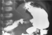
🔍 Figure 2 (Radiology): Contrast barium upper GI study (5-week-old infant with projectile vomiting). The image shows a dilated stomach, elongated narrowed pyloric channel with a thin linear column of barium ("string sign"), parallel double-track streaks ("double-track sign"), and a concave impression at the base of the duodenal bulb ("shoulder sign"). No contrast passes beyond the duodenal cap.
💡 Examiner instruction (interactive): The candidate will be asked to diagnose hypertrophic pyloric stenosis, describe the classic clinical triad (projectile vomiting, visible peristalsis, olive mass), interpret the barium radiograph (string sign, double-track sign, shoulder sign), order diagnostic studies (abdominal ultrasound is gold standard), discuss metabolic derangements (hypochloremic metabolic alkalosis), and outline treatment (IV fluid resuscitation followed by Ramstedt pyloromyotomy).
🔍 Examiner Questions (interactive) – Click to reveal model answers
❓ Q1 (Examiner): “What is the most likely diagnosis in this 5-week-old infant with projectile non-bilious vomiting, visible peristalsis, and an olive-shaped mass? List the classic clinical features of hypertrophic pyloric stenosis.”
✅ Candidate's answer:
• Diagnosis: Hypertrophic pyloric stenosis (HPS).
• Classic clinical features (triad):
1️⃣ Progressive, projectile, non-bilious vomiting typically starting at 4-6 weeks of age.
2️⃣ Visible gastric peristalsis (left-to-right wave across epigastrium).
3️⃣ Palpable olive-shaped mass in the right upper quadrant (most specific sign).
• Associated findings: Constant hunger, weight loss/failure to thrive, dehydration, hypochloremic metabolic alkalosis.
• Diagnosis: Hypertrophic pyloric stenosis (HPS).
• Classic clinical features (triad):
1️⃣ Progressive, projectile, non-bilious vomiting typically starting at 4-6 weeks of age.
2️⃣ Visible gastric peristalsis (left-to-right wave across epigastrium).
3️⃣ Palpable olive-shaped mass in the right upper quadrant (most specific sign).
• Associated findings: Constant hunger, weight loss/failure to thrive, dehydration, hypochloremic metabolic alkalosis.
❓ Q2 (Examiner): “What is the epidemiology of hypertrophic pyloric stenosis? Which infants are at highest risk?”
✅ Candidate's answer:
• Incidence: 2-5/1,000 live births (highest in Caucasians).
• M:F = 4:1 to 5:1. Risk factors: first-born male, family history, maternal smoking, bottle feeding, erythromycin exposure.
• Incidence: 2-5/1,000 live births (highest in Caucasians).
• M:F = 4:1 to 5:1. Risk factors: first-born male, family history, maternal smoking, bottle feeding, erythromycin exposure.
❓ Q3 (Examiner): “Explain the pathophysiology of hypertrophic pyloric stenosis.”
✅ Candidate's answer:
Idiopathic thickening of circular muscle layer of pylorus → progressive gastric outlet obstruction → gastric dilation, visible peristalsis, projectile vomiting. Histology: smooth muscle hypertrophy with decreased nitric oxide synthase-containing neurons.
Idiopathic thickening of circular muscle layer of pylorus → progressive gastric outlet obstruction → gastric dilation, visible peristalsis, projectile vomiting. Histology: smooth muscle hypertrophy with decreased nitric oxide synthase-containing neurons.
❓ Q4 (Examiner): “What electrolyte and acid-base disturbances occur? Explain paradoxical aciduria.”
✅ Candidate's answer:
Hypochloremic metabolic alkalosis with hypokalemia. Paradoxical aciduria: urine pH <5.5 despite systemic alkalosis due to volume depletion activating RAAS → Na+ retention, H+ excretion.
Hypochloremic metabolic alkalosis with hypokalemia. Paradoxical aciduria: urine pH <5.5 despite systemic alkalosis due to volume depletion activating RAAS → Na+ retention, H+ excretion.
❓ Q5 (Examiner): “What is the diagnostic imaging modality of choice? What are the criteria?”
✅ Candidate's answer:
Abdominal ultrasound (gold standard). Criteria: muscle thickness ≥4 mm, channel length ≥16 mm, pyloric diameter ≥10-12 mm.
Abdominal ultrasound (gold standard). Criteria: muscle thickness ≥4 mm, channel length ≥16 mm, pyloric diameter ≥10-12 mm.
❓ Q6 (Examiner): “What conditions should be considered in the differential diagnosis?”
✅ Candidate's answer:
GERD, malrotation with volvulus (bilious vomiting), duodenal atresia (double bubble), antral web, pylorospasm, milk protein allergy, intracranial pathology, metabolic disorders.
GERD, malrotation with volvulus (bilious vomiting), duodenal atresia (double bubble), antral web, pylorospasm, milk protein allergy, intracranial pathology, metabolic disorders.
❓ Q7 (Examiner): “How do you medically manage before surgery?”
✅ Candidate's answer:
IV normal saline + KCl (after urine output). Correct dehydration and alkalosis. NG decompression. Delay surgery until electrolytes normalized.
IV normal saline + KCl (after urine output). Correct dehydration and alkalosis. NG decompression. Delay surgery until electrolytes normalized.
❓ Q8 (Examiner): “What is the definitive surgical treatment? Describe the procedure.”
✅ Candidate's answer:
Ramstedt pyloromyotomy (open or laparoscopic). Longitudinal incision through serosa and circular muscle down to submucosa, allowing mucosa to bulge. Relieves obstruction.
Ramstedt pyloromyotomy (open or laparoscopic). Longitudinal incision through serosa and circular muscle down to submucosa, allowing mucosa to bulge. Relieves obstruction.
❓ Q9 (Examiner): “What are the findings on upper GI series in HPS?”
✅ Candidate's answer:
String sign (thin barium column), double-track sign (parallel streaks), shoulder sign (antral indentation), beak sign, delayed gastric emptying.
String sign (thin barium column), double-track sign (parallel streaks), shoulder sign (antral indentation), beak sign, delayed gastric emptying.
❓ Q10 (Examiner): “Is pyloromyotomy an emergency? When to operate?”
✅ Candidate's answer:
No, not an emergency. Surgery is semi-elective after correcting dehydration and alkalosis (usually 12-48 hours).
No, not an emergency. Surgery is semi-elective after correcting dehydration and alkalosis (usually 12-48 hours).
❓ Q11 (Examiner): “What are complications of pyloromyotomy?”
✅ Candidate's answer:
Mucosal perforation (1-2%), incomplete myotomy, bleeding, wound infection, postoperative vomiting (gastric atony).
Mucosal perforation (1-2%), incomplete myotomy, bleeding, wound infection, postoperative vomiting (gastric atony).
❓ Q12 (Examiner): “What is the prognosis after surgery?”
✅ Candidate's answer:
Excellent >95% resolution. Recurrence extremely rare (<0.5%). Normal long-term gastric function.
Excellent >95% resolution. Recurrence extremely rare (<0.5%). Normal long-term gastric function.
❓ Q13 (Examiner): “Is there any non-surgical treatment?”
✅ Candidate's answer:
IV atropine has been used but requires prolonged hospitalization, lower success rate, significant side effects. Pyloromyotomy remains gold standard.
IV atropine has been used but requires prolonged hospitalization, lower success rate, significant side effects. Pyloromyotomy remains gold standard.
❓ Q14 (Examiner): “How do you counsel the anxious parents?”
✅ Candidate's structured answer:
Explain condition (thickened pyloric muscle → obstruction). Reassure that it is treatable with surgery. Describe preoperative stabilization, the pyloromyotomy procedure, excellent prognosis, and typical hospital stay of 1-2 days.
Explain condition (thickened pyloric muscle → obstruction). Reassure that it is treatable with surgery. Describe preoperative stabilization, the pyloromyotomy procedure, excellent prognosis, and typical hospital stay of 1-2 days.
❓ Q15 (Examiner): “How does presentation differ in premature infants or those >3 months?”
✅ Candidate's answer:
Premature: later corrected age (40-44 weeks), less dramatic symptoms, higher postoperative apnea risk. Late >3 months: more weight loss, intermittent vomiting, consider alternate diagnoses.
Premature: later corrected age (40-44 weeks), less dramatic symptoms, higher postoperative apnea risk. Late >3 months: more weight loss, intermittent vomiting, consider alternate diagnoses.
❓ Q16 (Examiner - Radiology/Image Interpretation): “Look at Figure 2 (the contrast barium upper GI study in this 5-week-old infant with projectile vomiting). Describe the radiological findings in detail. Identify at least three specific signs seen on this barium study. What is the most likely diagnosis?”
✅ Candidate's structured answer (Image Interpretation):
1. Description of radiological findings:
• The stomach is markedly dilated (gastric distension) due to chronic outlet obstruction.
• There is delayed gastric emptying with minimal contrast passing into the duodenum.
• The pyloric channel is elongated and narrowed.
• Two parallel streaks of barium are seen within the pyloric channel → "double-track sign" (contrast trapped between thickened mucosal folds).
• A thin, linear column of contrast connects the antrum to the duodenum → "string sign" (pathognomonic).
• A concave filling defect is noted at the base of the duodenal bulb → "shoulder sign" (due to bulging of hypertrophied pyloric muscle).
• The proximal duodenal cap shows a small "beak" deformity.
2. Three specific signs (candidate must name at least three):
- String sign – thin, elongated, linear stream of barium through narrowed pyloric channel.
- Double-track sign – two parallel barium streaks within the pylorus (redundant mucosa).
- Shoulder sign – smooth indentation on the antrum from hypertrophied muscle.
- Beak sign – pointed tapering at pyloric entrance.
- Delayed gastric emptying – barium retention in stomach for >3 hours.
3. Most likely diagnosis:
• Hypertrophic Pyloric Stenosis (HPS)
4. Additional notes (examiner may probe):
• Gold standard is ultrasound (muscle thickness ≥4 mm, length ≥16 mm).
• Barium study is used if ultrasound equivocal or to exclude malrotation (bilious vomiting).
• Always correlate with electrolyte status (hypochloremic metabolic alkalosis).
• If bilious vomiting, suspect malrotation with volvulus (surgical emergency).
1. Description of radiological findings:
• The stomach is markedly dilated (gastric distension) due to chronic outlet obstruction.
• There is delayed gastric emptying with minimal contrast passing into the duodenum.
• The pyloric channel is elongated and narrowed.
• Two parallel streaks of barium are seen within the pyloric channel → "double-track sign" (contrast trapped between thickened mucosal folds).
• A thin, linear column of contrast connects the antrum to the duodenum → "string sign" (pathognomonic).
• A concave filling defect is noted at the base of the duodenal bulb → "shoulder sign" (due to bulging of hypertrophied pyloric muscle).
• The proximal duodenal cap shows a small "beak" deformity.
2. Three specific signs (candidate must name at least three):
- String sign – thin, elongated, linear stream of barium through narrowed pyloric channel.
- Double-track sign – two parallel barium streaks within the pylorus (redundant mucosa).
- Shoulder sign – smooth indentation on the antrum from hypertrophied muscle.
- Beak sign – pointed tapering at pyloric entrance.
- Delayed gastric emptying – barium retention in stomach for >3 hours.
3. Most likely diagnosis:
• Hypertrophic Pyloric Stenosis (HPS)
4. Additional notes (examiner may probe):
• Gold standard is ultrasound (muscle thickness ≥4 mm, length ≥16 mm).
• Barium study is used if ultrasound equivocal or to exclude malrotation (bilious vomiting).
• Always correlate with electrolyte status (hypochloremic metabolic alkalosis).
• If bilious vomiting, suspect malrotation with volvulus (surgical emergency).
🗣️ Examiner's probing / high-yield points:
• "What is the most specific physical finding?" → Palpable olive-shaped mass.
• "What are the diagnostic ultrasound criteria?" → Muscle thickness ≥4 mm, length ≥16 mm.
• "What is the classical electrolyte disturbance?" → Hypochloremic metabolic alkalosis with hypokalemia and paradoxical aciduria.
• "Why correct alkalosis before surgery?" → To prevent postoperative apnea and arrhythmias.
• "What is the definitive treatment?" → Ramstedt pyloromyotomy.
• "On barium study, which sign is considered most specific for HPS?" → String sign (though double-track and shoulder sign are also highly suggestive).
• "If this infant had bilious vomiting, what would you suspect?" → Malrotation with midgut volvulus.
• "What is the most specific physical finding?" → Palpable olive-shaped mass.
• "What are the diagnostic ultrasound criteria?" → Muscle thickness ≥4 mm, length ≥16 mm.
• "What is the classical electrolyte disturbance?" → Hypochloremic metabolic alkalosis with hypokalemia and paradoxical aciduria.
• "Why correct alkalosis before surgery?" → To prevent postoperative apnea and arrhythmias.
• "What is the definitive treatment?" → Ramstedt pyloromyotomy.
• "On barium study, which sign is considered most specific for HPS?" → String sign (though double-track and shoulder sign are also highly suggestive).
• "If this infant had bilious vomiting, what would you suspect?" → Malrotation with midgut volvulus.
📘 Hypertrophic Pyloric Stenosis – Core Revision for TOACS
🔍 Definition
Idiopathic hypertrophy of the circular pyloric muscle → gastric outlet obstruction. Incidence 2-5/1,000 live births. M:F 4:1. Peak at 4-6 weeks.
Idiopathic hypertrophy of the circular pyloric muscle → gastric outlet obstruction. Incidence 2-5/1,000 live births. M:F 4:1. Peak at 4-6 weeks.
🩺 Clinical Triad
1. Projectile non-bilious vomiting (hungry after vomiting)
2. Visible gastric peristalsis (left-to-right)
3. Palpable olive mass (RUQ) – most specific
1. Projectile non-bilious vomiting (hungry after vomiting)
2. Visible gastric peristalsis (left-to-right)
3. Palpable olive mass (RUQ) – most specific
🧪 Metabolic Derangements
Hypochloremic metabolic alkalosis, hypokalemia, paradoxical aciduria (urine pH <5.5). Prerenal azotemia.
Hypochloremic metabolic alkalosis, hypokalemia, paradoxical aciduria (urine pH <5.5). Prerenal azotemia.
📋 Diagnosis
Gold standard: Abdominal ultrasound – muscle thickness ≥4 mm, channel length ≥16 mm. Upper GI series (if equivocal): string sign, double-track sign, shoulder sign.
Gold standard: Abdominal ultrasound – muscle thickness ≥4 mm, channel length ≥16 mm. Upper GI series (if equivocal): string sign, double-track sign, shoulder sign.
💊 Preoperative Management
IV normal saline + KCl (after urine output). Correct dehydration and alkalosis before surgery. NG decompression.
IV normal saline + KCl (after urine output). Correct dehydration and alkalosis before surgery. NG decompression.
🔪 Surgical Treatment
Ramstedt pyloromyotomy (open or laparoscopic). Post-op feeds in 4-6 hours. Excellent prognosis; recurrence rare.
Ramstedt pyloromyotomy (open or laparoscopic). Post-op feeds in 4-6 hours. Excellent prognosis; recurrence rare.
⭐ High-yield pearls for TOACS (Hypertrophic Pyloric Stenosis):
• Typical presentation: 4-6 week old male with projectile non-bilious vomiting after feeds.
• Most specific sign: Olive-shaped mass (RUQ).
• Gold standard diagnostic test: Abdominal ultrasound (muscle thickness ≥4 mm).
• Classic electrolyte picture: Hypochloremic metabolic alkalosis + hypokalemia + paradoxical aciduria.
• Definitive treatment: Ramstedt pyloromyotomy (correct electrolytes first – not an emergency).
• Barium study signs: String sign (thin column), double-track sign (parallel streaks), shoulder sign (antral indentation).
• If bilious vomiting → suspect malrotation/volvulus.
• Typical presentation: 4-6 week old male with projectile non-bilious vomiting after feeds.
• Most specific sign: Olive-shaped mass (RUQ).
• Gold standard diagnostic test: Abdominal ultrasound (muscle thickness ≥4 mm).
• Classic electrolyte picture: Hypochloremic metabolic alkalosis + hypokalemia + paradoxical aciduria.
• Definitive treatment: Ramstedt pyloromyotomy (correct electrolytes first – not an emergency).
• Barium study signs: String sign (thin column), double-track sign (parallel streaks), shoulder sign (antral indentation).
• If bilious vomiting → suspect malrotation/volvulus.
🗣️ Candidate's role-play & examiner feedback
💬 To the candidate (role‑play): You will be asked the 16 questions from the Examiner Q&A tab (including clinical features, olive sign, ultrasound criteria, metabolic alkalosis, preoperative fluid management, Ramstedt pyloromyotomy, differential diagnosis, and radiology image interpretation of the barium study – string sign, double-track sign, shoulder sign). Provide concise, evidence‑based answers. Demonstrate empathy when counseling parents. Emphasize the importance of correcting dehydration and alkalosis before anesthesia.
📝 Examiner Marking Grid (Hypertrophic Pyloric Stenosis – TOACS station):
- ✅ Recognizes classic presentation (4-6 weeks, projectile non-bilious vomiting, hungry after vomiting)
- ✅ Identifies visible peristalsis and palpates olive mass (RUQ)
- ✅ Orders abdominal ultrasound as first-line diagnostic test
- ✅ Describes ultrasound criteria (muscle thickness ≥4 mm, length ≥16 mm)
- ✅ Identifies hypochloremic metabolic alkalosis with paradoxical aciduria
- ✅ Prescribes IV normal saline + KCl to correct dehydration/alkalosis
- ✅ Recommends Ramstedt pyloromyotomy after stabilization
- ✅ Interprets barium radiograph correctly (string sign, double-track sign, shoulder sign, delayed emptying)
- ✅ Provides appropriate parental counseling (excellent prognosis, surgery not an emergency)
- ✅ Lists differential diagnoses (GERD, malrotation, antral web)
📚 Key references: Nelson Textbook of Pediatrics 22e (Chapter 383 – Pyloric Stenosis), American Academy of Pediatrics guidelines, CPSP protocols for infantile vomiting, ESPGHAN guidelines.