FCPS Paediatrics TOACS · Interactive Station
🩺 Myelomeningocele (Spina Bifida Aperta) — Neural tube defect, prenatal diagnosis, surgical & long-term management
📚 Paeds Online – paeds.online
⚕️ OBSERVED STATION · CPSP FORMAT · 8 MINUTES · SEPARATE TABS · CLINICAL IMAGE INCLUDED
📖 Problem-oriented Clinical Scenario + Image
👶🏻 Clinical Scenario (read aloud – 2 min):
A 6-hour-old term female infant is referred to the neonatal unit from a peripheral nursery. The baby was born at 39 weeks via normal vaginal delivery to a 24-year-old primigravida mother. Antenatal history: mother did not take folic acid supplementation periconceptually; first trimester ultrasound was normal, but second trimester scan at 20 weeks showed a possible neural tube defect. On examination: The infant is alert and hemodynamically stable. There is a large, cystic, membrane-covered sac in the lumbosacral region (L4-S1) measuring ~5 cm. The sac is translucent with visible neural tissue placode. The lower limbs show flaccid paralysis, absent reflexes, and no withdrawal to pinprick. The anus is patulous, and there is dribbling of urine. Head circumference is at the 75th percentile. The infant has a high-arched palate and bilateral talipes equinovarus (clubfoot).
A clinical photograph of the lumbosacral lesion is shown below.
Task: Describe the findings, propose the most likely diagnosis, discuss immediate management, and answer the examiner's questions regarding etiology, associated anomalies, investigations, surgical timing, and long-term follow-up.
A 6-hour-old term female infant is referred to the neonatal unit from a peripheral nursery. The baby was born at 39 weeks via normal vaginal delivery to a 24-year-old primigravida mother. Antenatal history: mother did not take folic acid supplementation periconceptually; first trimester ultrasound was normal, but second trimester scan at 20 weeks showed a possible neural tube defect. On examination: The infant is alert and hemodynamically stable. There is a large, cystic, membrane-covered sac in the lumbosacral region (L4-S1) measuring ~5 cm. The sac is translucent with visible neural tissue placode. The lower limbs show flaccid paralysis, absent reflexes, and no withdrawal to pinprick. The anus is patulous, and there is dribbling of urine. Head circumference is at the 75th percentile. The infant has a high-arched palate and bilateral talipes equinovarus (clubfoot).
A clinical photograph of the lumbosacral lesion is shown below.
Task: Describe the findings, propose the most likely diagnosis, discuss immediate management, and answer the examiner's questions regarding etiology, associated anomalies, investigations, surgical timing, and long-term follow-up.
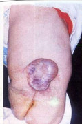
🔍 Figure: Lumbosacral myelomeningocele – a cystic, membrane-covered sac containing neural tissue (placode). This is the most severe form of spina bifida aperta, associated with neurological deficit below the level of the lesion.
Source: PostImages – Myelomeningocele.
Source: PostImages – Myelomeningocele.
💡 Examiner instruction (interactive): The candidate will be asked to identify the condition, interpret the image, describe associated anomalies (Chiari II, hydrocephalus, clubfoot), discuss prevention (folic acid), prenatal diagnosis, delivery room management (sterile dressing, positioning), surgical repair timing, MOMS trial results, and long-term multidisciplinary follow-up.
🔍 Examiner Questions (interactive) – Click to reveal model answers
❓ Q1 (Examiner): “Describe the findings in the image and give the most likely diagnosis. Differentiate from meningocele.”
✅ Candidate's structured answer:
• Findings: Lumbosacral cystic lesion, covered by thin membrane, visible neural placode (neural tissue) inside the sac. Surrounding skin is intact.
• Diagnosis: Myelomeningocele (spina bifida aperta) – the most severe form of open neural tube defect.
• Differentiation from meningocele: Meningocele contains only meninges and CSF, no neural tissue, and typically no neurological deficit. Myelomeningocele contains neural placode → motor/sensory deficit below the level.
• Findings: Lumbosacral cystic lesion, covered by thin membrane, visible neural placode (neural tissue) inside the sac. Surrounding skin is intact.
• Diagnosis: Myelomeningocele (spina bifida aperta) – the most severe form of open neural tube defect.
• Differentiation from meningocele: Meningocele contains only meninges and CSF, no neural tissue, and typically no neurological deficit. Myelomeningocele contains neural placode → motor/sensory deficit below the level.
❓ Q2 (Examiner): “What is the embryological basis and risk factors for myelomeningocele?”
✅ Candidate's answer:
• Embryology: Failure of neural tube closure during the 3rd-4th week of gestation (neurulation). Cranial end closes by day 24-26, caudal end (spinal) by day 28.
• Risk factors: Maternal folic acid deficiency (periconceptional), maternal diabetes, obesity, hyperthermia, valproate or carbamazepine use, family history (multifactorial inheritance; recurrence risk 3-5% after one affected child).
• Prevention: Periconceptional folic acid (400 mcg daily for general population; 4 mg daily for high-risk mothers with previous NTD-affected pregnancy).
• Embryology: Failure of neural tube closure during the 3rd-4th week of gestation (neurulation). Cranial end closes by day 24-26, caudal end (spinal) by day 28.
• Risk factors: Maternal folic acid deficiency (periconceptional), maternal diabetes, obesity, hyperthermia, valproate or carbamazepine use, family history (multifactorial inheritance; recurrence risk 3-5% after one affected child).
• Prevention: Periconceptional folic acid (400 mcg daily for general population; 4 mg daily for high-risk mothers with previous NTD-affected pregnancy).
❓ Q3 (Examiner): “What are the common associated congenital anomalies in myelomeningocele?”
✅ Candidate's answer:
• Chiari II malformation (hindbrain herniation) – almost always present, may cause brainstem compression, apnea, dysphagia.
• Hydrocephalus – occurs in 80-90% due to Chiari II obstructing CSF flow; often requires VP shunt.
• Orthopedic: Clubfoot (talipes equinovarus), hip dislocation, kyphoscoliosis.
• Neurogenic bladder – detrusor-sphincter dyssynergia, recurrent UTIs, renal scarring.
• Bowel dysfunction – neurogenic bowel with constipation/incontinence.
• Latex allergy – high incidence due to early surgical exposure.
• Chiari II malformation (hindbrain herniation) – almost always present, may cause brainstem compression, apnea, dysphagia.
• Hydrocephalus – occurs in 80-90% due to Chiari II obstructing CSF flow; often requires VP shunt.
• Orthopedic: Clubfoot (talipes equinovarus), hip dislocation, kyphoscoliosis.
• Neurogenic bladder – detrusor-sphincter dyssynergia, recurrent UTIs, renal scarring.
• Bowel dysfunction – neurogenic bowel with constipation/incontinence.
• Latex allergy – high incidence due to early surgical exposure.
❓ Q4 (Examiner): “How is myelomeningocele diagnosed prenatally?”
✅ Candidate's answer:
• Maternal serum alpha-fetoprotein (MSAFP) – elevated at 15-20 weeks (screening).
• Fetal ultrasound – shows "lemon sign" (bifrontal indentation) and "banana sign" (cerebellar herniation) along with spinal defect and sac.
• Amniotic fluid AFP and acetylcholinesterase (AChE) – elevated (diagnostic).
• Fetal MRI – detailed anatomy, brainstem, ventricular size, spinal cord level.
• Amniocentesis for karyotype/microarray – to rule out chromosomal anomalies (trisomy 18, 13).
• Maternal serum alpha-fetoprotein (MSAFP) – elevated at 15-20 weeks (screening).
• Fetal ultrasound – shows "lemon sign" (bifrontal indentation) and "banana sign" (cerebellar herniation) along with spinal defect and sac.
• Amniotic fluid AFP and acetylcholinesterase (AChE) – elevated (diagnostic).
• Fetal MRI – detailed anatomy, brainstem, ventricular size, spinal cord level.
• Amniocentesis for karyotype/microarray – to rule out chromosomal anomalies (trisomy 18, 13).
❓ Q5 (Examiner): “Describe the immediate postnatal management of this infant in the delivery room and NICU.”
✅ Candidate's answer:
1️⃣ Protect the sac – cover with sterile, saline-moistened non-adherent dressing; avoid drying or rupture. Do not apply antiseptics.
2️⃣ Positioning – prone or lateral to avoid pressure on the sac.
3️⃣ Assess for hydrocephalus – measure head circumference, fontanel, suture separation; serial cranial ultrasound.
4️⃣ Monitor for brainstem dysfunction – stridor, apnea, poor feeding, vocal cord paralysis (Chiari II).
5️⃣ Bladder management – monitor urine output, consider early urology referral and clean intermittent catheterization (CIC).
6️⃣ Antibiotics – start prophylactic IV antibiotics (e.g., ampicillin + gentamicin) until repair to prevent meningitis.
7️⃣ Latex precautions – use latex-free gloves/equipment from birth.
1️⃣ Protect the sac – cover with sterile, saline-moistened non-adherent dressing; avoid drying or rupture. Do not apply antiseptics.
2️⃣ Positioning – prone or lateral to avoid pressure on the sac.
3️⃣ Assess for hydrocephalus – measure head circumference, fontanel, suture separation; serial cranial ultrasound.
4️⃣ Monitor for brainstem dysfunction – stridor, apnea, poor feeding, vocal cord paralysis (Chiari II).
5️⃣ Bladder management – monitor urine output, consider early urology referral and clean intermittent catheterization (CIC).
6️⃣ Antibiotics – start prophylactic IV antibiotics (e.g., ampicillin + gentamicin) until repair to prevent meningitis.
7️⃣ Latex precautions – use latex-free gloves/equipment from birth.
❓ Q6 (Examiner): “When is surgical closure performed, and what are the principles of surgery?”
✅ Candidate's answer:
• Timing: Within 24-72 hours of birth (neonatal closure) to reduce risk of meningitis and further neurological injury.
• Principles: Excision of the sac, preservation of functional neural tissue, watertight dural closure, and skin coverage (may require rotational flaps or skin graft for large defects).
• Associated procedures: Ventriculoperitoneal (VP) shunt insertion may be done at same time or delayed depending on hydrocephalus severity.
• Postoperative: Continue prone positioning, monitor for CSF leak, infection, or wound dehiscence.
• Timing: Within 24-72 hours of birth (neonatal closure) to reduce risk of meningitis and further neurological injury.
• Principles: Excision of the sac, preservation of functional neural tissue, watertight dural closure, and skin coverage (may require rotational flaps or skin graft for large defects).
• Associated procedures: Ventriculoperitoneal (VP) shunt insertion may be done at same time or delayed depending on hydrocephalus severity.
• Postoperative: Continue prone positioning, monitor for CSF leak, infection, or wound dehiscence.
❓ Q7 (Examiner): “What is the role of fetal surgery in myelomeningocele? Summarize the MOMS trial results.”
✅ Candidate's answer:
• Fetal repair – open hysterotomy or fetoscopic closure performed before 26 weeks gestation in select centers.
• MOMS trial (Management of Myelomeningocele Study):
- Outcomes: Prenatal surgery reduced need for VP shunt at 12 months (40% vs 82%). Improved mental development and motor function at 30 months. Reduced hindbrain herniation.
- Risks: Increased preterm delivery, uterine dehiscence, and maternal complications.
• Candidate criteria: Gestational age 19-26 weeks, normal karyotype, lesion level T1-S1, no other major anomalies, maternal health.
• Fetal repair – open hysterotomy or fetoscopic closure performed before 26 weeks gestation in select centers.
• MOMS trial (Management of Myelomeningocele Study):
- Outcomes: Prenatal surgery reduced need for VP shunt at 12 months (40% vs 82%). Improved mental development and motor function at 30 months. Reduced hindbrain herniation.
- Risks: Increased preterm delivery, uterine dehiscence, and maternal complications.
• Candidate criteria: Gestational age 19-26 weeks, normal karyotype, lesion level T1-S1, no other major anomalies, maternal health.
❓ Q8 (Examiner): “How do you diagnose and manage hydrocephalus in a newborn with myelomeningocele?”
✅ Candidate's answer:
• Diagnosis: Serial head circumference measurement (crossing percentiles), bulging fontanel, suture separation, sunsetting sign. Cranial ultrasound to measure ventricular index (≥97th percentile for age) and assess for progressive ventriculomegaly.
• Management:
- Mild/stable ventriculomegaly: Observe; may stabilize without shunt.
- Progressive or symptomatic hydrocephalus: Ventriculoperitoneal (VP) shunt insertion (most common) or endoscopic third ventriculostomy (ETV) in select cases.
• Shunt complications: Infection, obstruction, overdrainage, need for revision (common, ~40% within first year).
• Diagnosis: Serial head circumference measurement (crossing percentiles), bulging fontanel, suture separation, sunsetting sign. Cranial ultrasound to measure ventricular index (≥97th percentile for age) and assess for progressive ventriculomegaly.
• Management:
- Mild/stable ventriculomegaly: Observe; may stabilize without shunt.
- Progressive or symptomatic hydrocephalus: Ventriculoperitoneal (VP) shunt insertion (most common) or endoscopic third ventriculostomy (ETV) in select cases.
• Shunt complications: Infection, obstruction, overdrainage, need for revision (common, ~40% within first year).
❓ Q9 (Examiner): “What is the long-term management of neurogenic bladder in myelomeningocele?”
✅ Candidate's answer:
• Early urology referral – baseline renal ultrasound, voiding cystourethrogram (VCUG), urodynamics.
• Clean intermittent catheterization (CIC) – started soon after birth to empty bladder, prevent hydronephrosis, UTIs.
• Anticholinergic medications – oxybutynin to reduce detrusor overactivity.
• Prophylactic antibiotics – controversial; used to prevent recurrent UTIs in some cases.
• Surgical options – Mitrofanoff (continent catheterizable stoma), bladder augmentation, or urinary diversion for refractory cases.
• Early urology referral – baseline renal ultrasound, voiding cystourethrogram (VCUG), urodynamics.
• Clean intermittent catheterization (CIC) – started soon after birth to empty bladder, prevent hydronephrosis, UTIs.
• Anticholinergic medications – oxybutynin to reduce detrusor overactivity.
• Prophylactic antibiotics – controversial; used to prevent recurrent UTIs in some cases.
• Surgical options – Mitrofanoff (continent catheterizable stoma), bladder augmentation, or urinary diversion for refractory cases.
❓ Q10 (Examiner): “How do you manage neurogenic bowel in these patients?”
✅ Candidate's answer:
• Constipation management: High-fiber diet, adequate hydration, stool softeners (polyethylene glycol), stimulants (senna).
• Bowel program: Daily suppositories or enemas to achieve scheduled bowel emptying.
• Transanal irrigation – Peristeen system for refractory constipation/fecal incontinence.
• Surgical options: Malone antegrade continence enema (MACE) – appendicostomy or cecostomy for antegrade enemas.
• Goal: Social continence (no accidents between scheduled bowel movements) by school age.
• Constipation management: High-fiber diet, adequate hydration, stool softeners (polyethylene glycol), stimulants (senna).
• Bowel program: Daily suppositories or enemas to achieve scheduled bowel emptying.
• Transanal irrigation – Peristeen system for refractory constipation/fecal incontinence.
• Surgical options: Malone antegrade continence enema (MACE) – appendicostomy or cecostomy for antegrade enemas.
• Goal: Social continence (no accidents between scheduled bowel movements) by school age.
❓ Q11 (Examiner): “What orthopedic issues are commonly seen and how are they managed?”
✅ Candidate's answer:
• Clubfoot (talipes equinovarus) – Ponseti method (serial casting) starting soon after birth, Achilles tenotomy if needed.
• Hip dislocation/subluxation – Observation initially; surgery if painful or causing functional impairment.
• Kyphoscoliosis – Monitor with X-rays; bracing; surgical correction for severe progressive curves.
• Lower limb weakness/paralysis – Physical therapy, orthotics (AFOs, KAFOs), mobility aids (walker, wheelchair).
• Pressure ulcers – Prevention with appropriate seating, cushions, skin checks.
• Clubfoot (talipes equinovarus) – Ponseti method (serial casting) starting soon after birth, Achilles tenotomy if needed.
• Hip dislocation/subluxation – Observation initially; surgery if painful or causing functional impairment.
• Kyphoscoliosis – Monitor with X-rays; bracing; surgical correction for severe progressive curves.
• Lower limb weakness/paralysis – Physical therapy, orthotics (AFOs, KAFOs), mobility aids (walker, wheelchair).
• Pressure ulcers – Prevention with appropriate seating, cushions, skin checks.
❓ Q12 (Examiner): “What long-term follow-up is required for a child with myelomeningocele?”
✅ Candidate's answer:
• Multidisciplinary team: Neurosurgery, urology, orthopedics, physical therapy, occupational therapy, developmental pediatrics, social work.
• Neurosurgical: Monitor for shunt malfunction, Chiari symptoms, tethered cord release if symptomatic.
• Urologic: Annual renal ultrasound, VCUG, urodynamics; monitor for UTIs, renal scarring.
• Developmental: Assess cognitive function (normal IQ in most but learning disabilities common), school support, early intervention.
• Latex allergy: Lifelong latex precautions; educate family and school.
• Transition to adult care: Planned transition around age 18-21 years.
• Multidisciplinary team: Neurosurgery, urology, orthopedics, physical therapy, occupational therapy, developmental pediatrics, social work.
• Neurosurgical: Monitor for shunt malfunction, Chiari symptoms, tethered cord release if symptomatic.
• Urologic: Annual renal ultrasound, VCUG, urodynamics; monitor for UTIs, renal scarring.
• Developmental: Assess cognitive function (normal IQ in most but learning disabilities common), school support, early intervention.
• Latex allergy: Lifelong latex precautions; educate family and school.
• Transition to adult care: Planned transition around age 18-21 years.
❓ Q13 (Examiner): “What is the prognosis? How will you counsel the parents about survival and quality of life?”
✅ Candidate's answer:
• Survival: With modern care, >85% survive to adulthood. Mortality highest in first few years (Chiari-related apnea, shunt complications, sepsis).
• Cognitive outcome: Most have normal intelligence (~70-80%), but specific learning disabilities (executive function, attention, math) are common. Hydrocephalus and shunt infections increase risk of cognitive impairment.
• Mobility: Depends on lesion level. Lumbar/sacral lesions may ambulate with orthotics; thoracic lesions typically wheelchair-dependent.
• Continence: Most achieve social continence of bowel and bladder with CIC and bowel program.
• Counseling points: Recurrence risk for next pregnancy is 3-5% (increased to 10-15% after two affected children). Recommend high-dose folic acid (4 mg/day) preconceptually. Emphasize multidisciplinary care and excellent functional outcomes.
• Survival: With modern care, >85% survive to adulthood. Mortality highest in first few years (Chiari-related apnea, shunt complications, sepsis).
• Cognitive outcome: Most have normal intelligence (~70-80%), but specific learning disabilities (executive function, attention, math) are common. Hydrocephalus and shunt infections increase risk of cognitive impairment.
• Mobility: Depends on lesion level. Lumbar/sacral lesions may ambulate with orthotics; thoracic lesions typically wheelchair-dependent.
• Continence: Most achieve social continence of bowel and bladder with CIC and bowel program.
• Counseling points: Recurrence risk for next pregnancy is 3-5% (increased to 10-15% after two affected children). Recommend high-dose folic acid (4 mg/day) preconceptually. Emphasize multidisciplinary care and excellent functional outcomes.
❓ Q14 (Examiner): “Why is latex allergy common in myelomeningocele, and how is it prevented?”
✅ Candidate's answer:
• Incidence: Up to 30-50% of children with myelomeningocele develop latex allergy due to repeated surgical and medical exposures (catheters, gloves, equipment).
• Prevention: Use latex-free gloves, catheters, and equipment from birth. Label chart prominently. Avoid latex balloons, pacifiers, and toys in hospital and home.
• Signs of reaction: Contact urticaria, rhinoconjunctivitis, anaphylaxis during surgery or catheterization. Refer to allergy specialist.
• Preoperative protocol: Latex-free operating room, schedule as first case of day to minimize environmental latex.
• Incidence: Up to 30-50% of children with myelomeningocele develop latex allergy due to repeated surgical and medical exposures (catheters, gloves, equipment).
• Prevention: Use latex-free gloves, catheters, and equipment from birth. Label chart prominently. Avoid latex balloons, pacifiers, and toys in hospital and home.
• Signs of reaction: Contact urticaria, rhinoconjunctivitis, anaphylaxis during surgery or catheterization. Refer to allergy specialist.
• Preoperative protocol: Latex-free operating room, schedule as first case of day to minimize environmental latex.
🗣️ Examiner's probing / high-yield points:
• "What is the difference between myelomeningocele and meningocele?" → Myelomeningocele contains neural placode with neurological deficit; meningocele has only meninges/CSF, normal neurology.
• "What are the lemon and banana signs on prenatal ultrasound?" → Lemon sign: bifrontal indentation; banana sign: cerebellar herniation with obliterated cisterna magna. Both indicate Chiari II malformation.
• "What is the Chiari II malformation?" → Downward displacement of cerebellar vermis, medulla, and fourth ventricle through foramen magnum, causing brainstem compression and hydrocephalus.
• "What is the recurrence risk for NTDs?" → 3-5% after one affected child; 10-15% after two affected. All future pregnancies require high-dose folic acid (4 mg/day) starting 1 month preconception.
• "Why is the 'prone position' important postnatally?" → To avoid pressure on the myelomeningocele sac, prevent rupture, and reduce risk of meningitis and neurological injury.
• "What is the difference between myelomeningocele and meningocele?" → Myelomeningocele contains neural placode with neurological deficit; meningocele has only meninges/CSF, normal neurology.
• "What are the lemon and banana signs on prenatal ultrasound?" → Lemon sign: bifrontal indentation; banana sign: cerebellar herniation with obliterated cisterna magna. Both indicate Chiari II malformation.
• "What is the Chiari II malformation?" → Downward displacement of cerebellar vermis, medulla, and fourth ventricle through foramen magnum, causing brainstem compression and hydrocephalus.
• "What is the recurrence risk for NTDs?" → 3-5% after one affected child; 10-15% after two affected. All future pregnancies require high-dose folic acid (4 mg/day) starting 1 month preconception.
• "Why is the 'prone position' important postnatally?" → To avoid pressure on the myelomeningocele sac, prevent rupture, and reduce risk of meningitis and neurological injury.
📘 Myelomeningocele (Spina Bifida Aperta) – Core Revision for TOACS
🔍 Definition
Neural tube defect (failure of caudal neurulation). Open defect with neural placode → motor/sensory deficit below the lesion level.
Neural tube defect (failure of caudal neurulation). Open defect with neural placode → motor/sensory deficit below the lesion level.
🧬 Prevention
Periconceptional folic acid (400 mcg/day routine; 4 mg/day high-risk). Reduces NTD incidence by 70%.
Periconceptional folic acid (400 mcg/day routine; 4 mg/day high-risk). Reduces NTD incidence by 70%.
📊 Associated anomalies
Chiari II malformation (universal), hydrocephalus (80-90%), neurogenic bladder, clubfoot, hip dislocation, latex allergy.
Chiari II malformation (universal), hydrocephalus (80-90%), neurogenic bladder, clubfoot, hip dislocation, latex allergy.
🩺 Prenatal Dx
Elevated MSAFP + amniotic fluid AChE; ultrasound: lemon sign, banana sign, spinal defect.
Elevated MSAFP + amniotic fluid AChE; ultrasound: lemon sign, banana sign, spinal defect.
⚕️ Fetal surgery (MOMS)
Prenatal closure (19-26 weeks) reduces shunt rate (40% vs 82%) and improves motor outcomes, but increases prematurity and maternal risks.
Prenatal closure (19-26 weeks) reduces shunt rate (40% vs 82%) and improves motor outcomes, but increases prematurity and maternal risks.
📈 Postnatal management
Protect sac (sterile moist dressing), latex precautions, prophylactic antibiotics, closure within 24-72h. Multidisciplinary long-term follow-up.
Protect sac (sterile moist dressing), latex precautions, prophylactic antibiotics, closure within 24-72h. Multidisciplinary long-term follow-up.
⭐ High-yield pearls for TOACS (Myelomeningocele):
• Clinical hallmark: Sac containing neural placode with lower limb flaccid paralysis, bladder/bowel dysfunction, and associated Chiari II/hydrocephalus.
• Immediate priorities: Cover sac (sterile, moist, non-adherent), prone positioning, latex-free environment, IV antibiotics.
• MOMS trial key message: Fetal repair significantly reduces need for VP shunt and improves motor outcomes but carries maternal/fetal risks.
• Long-term multidisciplinary team: Neurosurgery, urology, orthopedics, PT/OT, developmental pediatrics, social work.
• Counseling pearls: Recurrence risk 3-5%; high-dose folic acid for next pregnancy. Normal intelligence expected in most.
• Clinical hallmark: Sac containing neural placode with lower limb flaccid paralysis, bladder/bowel dysfunction, and associated Chiari II/hydrocephalus.
• Immediate priorities: Cover sac (sterile, moist, non-adherent), prone positioning, latex-free environment, IV antibiotics.
• MOMS trial key message: Fetal repair significantly reduces need for VP shunt and improves motor outcomes but carries maternal/fetal risks.
• Long-term multidisciplinary team: Neurosurgery, urology, orthopedics, PT/OT, developmental pediatrics, social work.
• Counseling pearls: Recurrence risk 3-5%; high-dose folic acid for next pregnancy. Normal intelligence expected in most.
🗣️ Candidate's role-play & examiner feedback
💬 To the candidate (role‑play): You will be asked the 14 questions from the Examiner Q&A tab (including image identification, differentials, prenatal diagnosis, management of hydrocephalus and neurogenic bladder, fetal surgery, and long-term follow-up). Provide concise, evidence‑based answers. Examiner may ask for clarification on the MOMS trial or Chiari complications. Use structured points and demonstrate empathy when counseling parents.
📝 Examiner Marking Grid (Myelomeningocele – TOACS station):
- ✅ Correctly identifies the image as myelomeningocele (differentiates from meningocele)
- ✅ Explains embryology and risk factors (folic acid deficiency, valproate, diabetes)
- ✅ Lists associated anomalies (Chiari II, hydrocephalus, clubfoot, neurogenic bladder, latex allergy)
- ✅ Describes prenatal diagnosis (MSAFP, US lemon/banana signs, amniotic AChE)
- ✅ Outlines immediate postnatal care (protect sac, latex precautions, antibiotics, positioning)
- ✅ Knows timing of surgical closure (24-72 hours) and principles
- ✅ Summarizes MOMS trial findings (reduced shunt, improved motor outcomes, risks)
- ✅ Demonstrates knowledge of neurogenic bladder management (CIC, anticholinergics, urology follow-up)
- ✅ Discusses long-term multidisciplinary follow-up and transition to adult care
- ✅ Provides appropriate parental counseling (recurrence risk, folic acid, prognosis)
📚 Key references: Nelson Textbook of Pediatrics 22e (Neural Tube Defects), MOMS trial (NEJM 2011), CPSP guidelines on NTD prevention, American Academy of Pediatrics (Spina Bifida clinical report).