FCPS Paediatrics TOACS · Interactive Station
🩺 Port-Wine Stain (Nevus Flammeus) — Capillary malformation, Sturge-Weber syndrome, glaucoma risk, laser therapy
📚 Paeds Online – paeds.online
⚕️ OBSERVED STATION · CPSP FORMAT · 8 MINUTES · SEPARATE TABS · CLINICAL PHOTO INCLUDED
📖 Problem-oriented Clinical Scenario + Photograph
👶🏻 Clinical Scenario (read aloud – 2 min):
A 1-day-old term female infant is examined in the newborn nursery. The mother reports a flat, pink to dark-red mark on the right side of the face noted since birth. It does not blanch completely with pressure and is not elevated. The mark involves the right forehead, upper eyelid, and malar area (V1 and V2 distribution of trigeminal nerve). The rest of the physical examination is normal, including the neurological exam. The mother is worried and asks what this mark is, whether it will go away, and if it is dangerous.
A clinical photograph of the facial lesion is shown below.
Task: Describe the findings, propose the most likely diagnosis, differentiate from other newborn vascular lesions, discuss associated syndromes (Sturge-Weber syndrome, glaucoma), and outline management and parental counseling.
A 1-day-old term female infant is examined in the newborn nursery. The mother reports a flat, pink to dark-red mark on the right side of the face noted since birth. It does not blanch completely with pressure and is not elevated. The mark involves the right forehead, upper eyelid, and malar area (V1 and V2 distribution of trigeminal nerve). The rest of the physical examination is normal, including the neurological exam. The mother is worried and asks what this mark is, whether it will go away, and if it is dangerous.
A clinical photograph of the facial lesion is shown below.
Task: Describe the findings, propose the most likely diagnosis, differentiate from other newborn vascular lesions, discuss associated syndromes (Sturge-Weber syndrome, glaucoma), and outline management and parental counseling.
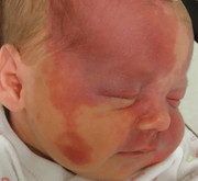
🔍 Figure: Newborn with a port-wine stain (nevus flammeus) on the right face, involving the forehead, upper eyelid, and malar region (V1 and V2 dermatomes). This distribution warrants evaluation for Sturge-Weber syndrome (leptomeningeal angiomatosis) and glaucoma.
💡 Examiner instruction (interactive): The candidate will be asked to identify port-wine stain (capillary malformation), differentiate from salmon patch (stork bite) and infantile hemangioma, discuss associations with Sturge-Weber syndrome (neurological) and glaucoma (ophthalmological), recommend appropriate screening (MRI brain, ophthalmology exam), and outline treatment (pulsed dye laser) and parental reassurance.
🔍 Examiner Questions (interactive) – Click to reveal model answers
❓ Q1 (Examiner): “Describe the findings in the image. What is the most likely diagnosis?”
✅ Candidate's structured answer:
• Findings: Flat, well-demarcated, pink to dark-red patch on the right face, involving forehead, upper eyelid, and malar area. Non-blanching (or incompletely blanching), not elevated, present since birth.
• Diagnosis: Port-wine stain (nevus flammeus) – a congenital capillary malformation.
• Findings: Flat, well-demarcated, pink to dark-red patch on the right face, involving forehead, upper eyelid, and malar area. Non-blanching (or incompletely blanching), not elevated, present since birth.
• Diagnosis: Port-wine stain (nevus flammeus) – a congenital capillary malformation.
❓ Q2 (Examiner): “What is the pathophysiology of a port-wine stain? How does it differ from a salmon patch (stork bite)?”
✅ Candidate's answer:
• Port-wine stain: Capillary malformation due to progressive dilatation of mature dermal capillaries and post-capillary venules. Results from somatic mutations in GNAQ gene (c.548G→A). Present at birth, does not involute, and may thicken/darken with age.
• Salmon patch (nevus simplex, "stork bite"): Also a capillary malformation but involves different vessels (more superficial). Usually on glabella, eyelids, or nape of neck. Fades or resolves completely by 1-2 years of age. No association with Sturge-Weber syndrome.
• Port-wine stains persist and may cause complications; salmon patches are benign.
• Port-wine stain: Capillary malformation due to progressive dilatation of mature dermal capillaries and post-capillary venules. Results from somatic mutations in GNAQ gene (c.548G→A). Present at birth, does not involute, and may thicken/darken with age.
• Salmon patch (nevus simplex, "stork bite"): Also a capillary malformation but involves different vessels (more superficial). Usually on glabella, eyelids, or nape of neck. Fades or resolves completely by 1-2 years of age. No association with Sturge-Weber syndrome.
• Port-wine stains persist and may cause complications; salmon patches are benign.
❓ Q3 (Examiner): “What are the differential diagnoses of a congenital red/purple facial lesion in a newborn?”
✅ Candidate's answer:
• Port-wine stain (nevus flammeus) – persistent, dark, V1/V2 distribution.
• Salmon patch (nevus simplex, stork bite) – fades, glabella/eyelids/nape.
• Infantile hemangioma – appears in first weeks, proliferative phase (grows), then involutes. Elevated, rubbery, may ulcerate.
• Cutaneous mastocytosis (urticaria pigmentosa) – red-brown macules, Darier sign (urticates with rubbing).
• Capillary malformation in other syndromes (CM-AVM, RASA1 mutations).
• Birth trauma (ecchymosis) – follows traumatic delivery, resolves in days.
• Key distinguishing feature: Port-wine stain does not blanch completely, does not regress, and may be associated with syndromes.
• Port-wine stain (nevus flammeus) – persistent, dark, V1/V2 distribution.
• Salmon patch (nevus simplex, stork bite) – fades, glabella/eyelids/nape.
• Infantile hemangioma – appears in first weeks, proliferative phase (grows), then involutes. Elevated, rubbery, may ulcerate.
• Cutaneous mastocytosis (urticaria pigmentosa) – red-brown macules, Darier sign (urticates with rubbing).
• Capillary malformation in other syndromes (CM-AVM, RASA1 mutations).
• Birth trauma (ecchymosis) – follows traumatic delivery, resolves in days.
• Key distinguishing feature: Port-wine stain does not blanch completely, does not regress, and may be associated with syndromes.
❓ Q4 (Examiner): “What is Sturge-Weber syndrome? Which port-wine stain location is most concerning for SWS?”
✅ Candidate's answer:
• Sturge-Weber syndrome (encephalotrigeminal angiomatosis): Sporadic neurocutaneous disorder characterized by leptomeningeal angiomatosis (pia mater), ipsilateral facial port-wine stain (V1 distribution), and glaucoma.
• High-risk location: Port-wine stain involving V1 (ophthalmic) branch of trigeminal nerve (forehead, upper eyelid, periorbital region). Risk is highest when both V1 and V2 are involved (as in this infant).
• Risk: Approximately 15-30% of infants with V1 port-wine stain have SWS. Bilateral or extensive lesions have higher risk.
• Neurological manifestations: Seizures (often focal, onset before 1 year), hemiparesis, developmental delay, intellectual disability, visual field defects.
• Sturge-Weber syndrome (encephalotrigeminal angiomatosis): Sporadic neurocutaneous disorder characterized by leptomeningeal angiomatosis (pia mater), ipsilateral facial port-wine stain (V1 distribution), and glaucoma.
• High-risk location: Port-wine stain involving V1 (ophthalmic) branch of trigeminal nerve (forehead, upper eyelid, periorbital region). Risk is highest when both V1 and V2 are involved (as in this infant).
• Risk: Approximately 15-30% of infants with V1 port-wine stain have SWS. Bilateral or extensive lesions have higher risk.
• Neurological manifestations: Seizures (often focal, onset before 1 year), hemiparesis, developmental delay, intellectual disability, visual field defects.
❓ Q5 (Examiner): “What is the risk of glaucoma with periorbital/eyelid port-wine stain? How is it screened?”
✅ Candidate's answer:
• Risk: Ipsilateral glaucoma occurs in ~30-50% of patients with port-wine stain involving the eyelid and V1 distribution. Can be congenital (present at birth) or develop in childhood.
• Glaucoma is due to elevated episcleral venous pressure and/or anterior chamber malformations.
• Screening:
- Ophthalmology examination at diagnosis (in newborn period).
- Repeat exams every 3-6 months in first year, then annually – lifelong risk, but highest in first decade.
- Measure intraocular pressure (IOP), optic disc cupping, corneal diameter, refraction.
• Treatment: Medical (beta-blockers, prostaglandin analogs) or surgical (goniotomy, trabeculectomy, drainage implants) for elevated IOP.
• Risk: Ipsilateral glaucoma occurs in ~30-50% of patients with port-wine stain involving the eyelid and V1 distribution. Can be congenital (present at birth) or develop in childhood.
• Glaucoma is due to elevated episcleral venous pressure and/or anterior chamber malformations.
• Screening:
- Ophthalmology examination at diagnosis (in newborn period).
- Repeat exams every 3-6 months in first year, then annually – lifelong risk, but highest in first decade.
- Measure intraocular pressure (IOP), optic disc cupping, corneal diameter, refraction.
• Treatment: Medical (beta-blockers, prostaglandin analogs) or surgical (goniotomy, trabeculectomy, drainage implants) for elevated IOP.
❓ Q6 (Examiner): “What neurological investigations are indicated for an infant with V1 port-wine stain?”
✅ Candidate's answer:
• Brain MRI with contrast (gadolinium) – imaging of choice to detect leptomeningeal angiomatosis (pial enhancement, usually occipital/parietal lobes ipsilateral to stain). Also assess for choroid plexus enlargement, cerebral atrophy, tram-track calcifications (seen later on CT).
• MRI timing: Asymptomatic infant – usually performed within first 6-12 months (some centers at 6 months). Earlier if neurological symptoms (seizures, hemiparesis).
• EEG: Not diagnostic but may show focal slowing or epileptiform discharges if SWS present; helps characterize seizure focus.
• Neurology referral: For baseline assessment and follow-up.
• If MRI negative but SWS suspected: Repeat MRI after 1-2 years (angiomatosis may become more evident).
• Brain MRI with contrast (gadolinium) – imaging of choice to detect leptomeningeal angiomatosis (pial enhancement, usually occipital/parietal lobes ipsilateral to stain). Also assess for choroid plexus enlargement, cerebral atrophy, tram-track calcifications (seen later on CT).
• MRI timing: Asymptomatic infant – usually performed within first 6-12 months (some centers at 6 months). Earlier if neurological symptoms (seizures, hemiparesis).
• EEG: Not diagnostic but may show focal slowing or epileptiform discharges if SWS present; helps characterize seizure focus.
• Neurology referral: For baseline assessment and follow-up.
• If MRI negative but SWS suspected: Repeat MRI after 1-2 years (angiomatosis may become more evident).
❓ Q7 (Examiner): “How would you manage a neonate with Sturge-Weber syndrome and seizures?”
✅ Candidate's answer:
• First-line anticonvulsants: Levetiracetam or oxcarbazepine for focal seizures. Phenobarbital may be used but has cognitive side effects.
• Prompt control of seizures – early, aggressive treatment may reduce neurodevelopmental impairment.
• Low-dose aspirin (antiplatelet) – considered by some to reduce risk of strokes/transient ischemic attacks (controversial, but widely used in SWS to improve perfusion).
• Vagus nerve stimulation or epilepsy surgery (focal resection, hemispherectomy) for refractory seizures.
• Multidisciplinary care: Neurology, ophthalmology, dermatology, developmental pediatrics.
• Parental counseling: Education on seizure recognition and emergency management.
• First-line anticonvulsants: Levetiracetam or oxcarbazepine for focal seizures. Phenobarbital may be used but has cognitive side effects.
• Prompt control of seizures – early, aggressive treatment may reduce neurodevelopmental impairment.
• Low-dose aspirin (antiplatelet) – considered by some to reduce risk of strokes/transient ischemic attacks (controversial, but widely used in SWS to improve perfusion).
• Vagus nerve stimulation or epilepsy surgery (focal resection, hemispherectomy) for refractory seizures.
• Multidisciplinary care: Neurology, ophthalmology, dermatology, developmental pediatrics.
• Parental counseling: Education on seizure recognition and emergency management.
❓ Q8 (Examiner): “What is the treatment for port-wine stain? When is it initiated?”
✅ Candidate's answer:
• First-line: Pulsed dye laser (PDL) – targets oxyhemoglobin in dilated vessels.
• Goal: Lighten the stain, prevent thickening/nodularity, improve cosmesis. Complete clearance is rare; significant lightening occurs in 50-80%.
• Timing: Can be started in infancy (as early as 2-3 months). Early treatment (before 1 year) yields better results and may prevent psychosocial impact.
• Procedure: Multiple sessions (every 4-8 weeks) under topical or general anesthesia (for extensive or facial lesions).
• Side effects: Temporary purpura, swelling, crusting, transient hyperpigmentation/hypopigmentation, scarring (rare).
• Parental counseling: Treatment is cosmetic, not medically necessary for the skin lesion itself, but recommended to prevent disfigurement and psychosocial distress.
• First-line: Pulsed dye laser (PDL) – targets oxyhemoglobin in dilated vessels.
• Goal: Lighten the stain, prevent thickening/nodularity, improve cosmesis. Complete clearance is rare; significant lightening occurs in 50-80%.
• Timing: Can be started in infancy (as early as 2-3 months). Early treatment (before 1 year) yields better results and may prevent psychosocial impact.
• Procedure: Multiple sessions (every 4-8 weeks) under topical or general anesthesia (for extensive or facial lesions).
• Side effects: Temporary purpura, swelling, crusting, transient hyperpigmentation/hypopigmentation, scarring (rare).
• Parental counseling: Treatment is cosmetic, not medically necessary for the skin lesion itself, but recommended to prevent disfigurement and psychosocial distress.
❓ Q9 (Examiner): “What other complications can occur with port-wine stains, especially on the extremities?”
✅ Candidate's answer:
• Soft tissue and bony hypertrophy – limb enlargement (hemihypertrophy or macrocheilia, macroglossia in facial lesions).
• Vascular blebs/nodules – progressive development of raised, bleeding nodules (pyogenic granuloma-like) in adulthood.
• Pain and functional impairment – if lesion involves joint areas or causes chronic swelling.
• Capillary malformation-arteriovenous malformation (CM-AVM) syndrome: when multiple capillary malformations + fast-flow lesions (need MRI/MRA).
• Coagulopathy (Kasabach-Merritt phenomenon) – does NOT occur with simple port-wine stains (only with kaposiform hemangioendothelioma/tufted angioma).
• Management: Orthopedic evaluation, compression garments for limb hypertrophy, laser for superficial lesions.
• Soft tissue and bony hypertrophy – limb enlargement (hemihypertrophy or macrocheilia, macroglossia in facial lesions).
• Vascular blebs/nodules – progressive development of raised, bleeding nodules (pyogenic granuloma-like) in adulthood.
• Pain and functional impairment – if lesion involves joint areas or causes chronic swelling.
• Capillary malformation-arteriovenous malformation (CM-AVM) syndrome: when multiple capillary malformations + fast-flow lesions (need MRI/MRA).
• Coagulopathy (Kasabach-Merritt phenomenon) – does NOT occur with simple port-wine stains (only with kaposiform hemangioendothelioma/tufted angioma).
• Management: Orthopedic evaluation, compression garments for limb hypertrophy, laser for superficial lesions.
❓ Q10 (Examiner): “What is the natural history of an untreated port-wine stain?”
✅ Candidate's answer:
• Does not resolve spontaneously. Persists throughout life.
• Color changes: Darkens from pink to dark purple/red over years.
• Texture changes: Becomes thick, cobblestoned, and nodular (bleeding nodules) by adulthood (40-50% by age 50).
• Progressive soft tissue hypertrophy (lips, gingiva, underlying bone).
• Psychosocial impact: Facial lesions cause significant social stigma, anxiety, depression.
• Syndromic complications: Sturge-Weber (seizures, hemiparesis, developmental delay) and glaucoma manifest early in childhood if present.
• Early pulsed dye laser treatment modifies the course (reduces thickening, lightens color).
• Does not resolve spontaneously. Persists throughout life.
• Color changes: Darkens from pink to dark purple/red over years.
• Texture changes: Becomes thick, cobblestoned, and nodular (bleeding nodules) by adulthood (40-50% by age 50).
• Progressive soft tissue hypertrophy (lips, gingiva, underlying bone).
• Psychosocial impact: Facial lesions cause significant social stigma, anxiety, depression.
• Syndromic complications: Sturge-Weber (seizures, hemiparesis, developmental delay) and glaucoma manifest early in childhood if present.
• Early pulsed dye laser treatment modifies the course (reduces thickening, lightens color).
❓ Q11 (Examiner): “How will you counsel the mother of this newborn with a V1/V2 port-wine stain?”
✅ Candidate's structured answer:
• “This is a port-wine stain – a collection of widened blood vessels in the skin. It is not cancerous and does not hurt.”
• “Unlike a stork bite, this mark will not go away on its own. It may darken and thicken over time, but it can be treated with a laser (pulsed dye laser) to make it much lighter.”
• “Because the mark is on the forehead and eyelid (V1 area), there is a small chance (15-30%) of Sturge-Weber syndrome – a condition with blood vessel changes on the brain surface that can cause seizures or weakness. We will arrange an MRI of the brain to check for this.”
• “There is also a risk of glaucoma (high pressure in the eye) on the same side as the stain. An eye doctor (ophthalmologist) will need to examine the baby regularly – first now, then every 3-6 months.”
• “Most children with port-wine stains have completely normal development, especially if the MRI is normal. Early treatment helps prevent complications.”
• “We will connect you with dermatology (for laser), ophthalmology, and neurology for coordinated care.”
• “This is a port-wine stain – a collection of widened blood vessels in the skin. It is not cancerous and does not hurt.”
• “Unlike a stork bite, this mark will not go away on its own. It may darken and thicken over time, but it can be treated with a laser (pulsed dye laser) to make it much lighter.”
• “Because the mark is on the forehead and eyelid (V1 area), there is a small chance (15-30%) of Sturge-Weber syndrome – a condition with blood vessel changes on the brain surface that can cause seizures or weakness. We will arrange an MRI of the brain to check for this.”
• “There is also a risk of glaucoma (high pressure in the eye) on the same side as the stain. An eye doctor (ophthalmologist) will need to examine the baby regularly – first now, then every 3-6 months.”
• “Most children with port-wine stains have completely normal development, especially if the MRI is normal. Early treatment helps prevent complications.”
• “We will connect you with dermatology (for laser), ophthalmology, and neurology for coordinated care.”
❓ Q12 (Examiner): “Is port-wine stain hereditary? What is the genetic mutation?”
✅ Candidate's answer:
• Port-wine stains are not inherited. They are caused by a somatic mutation (post-zygotic) in the GNAQ gene (c.548G→A, p.R183Q) in the endothelial cells of the affected vessels.
• The mutation is not present in the germline, so it is not passed to offspring.
• Sturge-Weber syndrome is also caused by the same somatic GNAQ mutation, affecting neural crest-derived tissues (skin, leptomeninges, eye).
• Recurrence risk for future children: Extremely low (<1%). The condition is sporadic.
• Parental reassurance: “You did nothing to cause this; it is a random change in a single cell during development.”
• Port-wine stains are not inherited. They are caused by a somatic mutation (post-zygotic) in the GNAQ gene (c.548G→A, p.R183Q) in the endothelial cells of the affected vessels.
• The mutation is not present in the germline, so it is not passed to offspring.
• Sturge-Weber syndrome is also caused by the same somatic GNAQ mutation, affecting neural crest-derived tissues (skin, leptomeninges, eye).
• Recurrence risk for future children: Extremely low (<1%). The condition is sporadic.
• Parental reassurance: “You did nothing to cause this; it is a random change in a single cell during development.”
❓ Q13 (Examiner): “What red flags in a child with port-wine stain warrant urgent evaluation?”
✅ Candidate's answer:
• Neurological: New-onset seizures (especially focal), prolonged staring spells, hemiparesis (weakness one side), loss of developmental milestones, acute altered mental status.
• Ophthalmological: Eye redness, corneal enlargement, excessive tearing, photophobia, poor visual tracking, suspected elevated eye pressure (buphthalmos).
• Dermatological: Ulceration, bleeding from nodules, signs of infection.
• Systemic: High-output cardiac failure (very rare, only with extensive trunk/extremity lesions with AV shunting).
• Any of these requires immediate neurology/ophthalmology/emergency care.
• Neurological: New-onset seizures (especially focal), prolonged staring spells, hemiparesis (weakness one side), loss of developmental milestones, acute altered mental status.
• Ophthalmological: Eye redness, corneal enlargement, excessive tearing, photophobia, poor visual tracking, suspected elevated eye pressure (buphthalmos).
• Dermatological: Ulceration, bleeding from nodules, signs of infection.
• Systemic: High-output cardiac failure (very rare, only with extensive trunk/extremity lesions with AV shunting).
• Any of these requires immediate neurology/ophthalmology/emergency care.
❓ Q14 (Examiner): “What is the recommended long-term surveillance for an infant with an isolated V1 port-wine stain (no SWS on MRI, normal eye exam)?”
✅ Candidate's answer:
• Ophthalmology: Examination every 3-6 months for the first year, then annually until at least 10 years (glaucoma risk persists, though highest in first decade).
• Neurology: Annual developmental assessments; no routine MRI if baseline MRI is normal and no neurological symptoms. Repeat MRI only if new symptoms (seizures, hemiparesis, cognitive decline).
• Dermatology: Follow-up for pulsed dye laser treatments (typically multiple sessions every 4-8 weeks). Monitor for thickening/nodularity.
• Psychosocial: Monitor for bullying, self-esteem issues, anxiety. Consider referral to psychology/counseling if needed.
• General pediatric follow-up: Regular well-child visits; alert parents to signs of seizures or vision problems.
• No activity restrictions. Normal childhood development expected.
• Ophthalmology: Examination every 3-6 months for the first year, then annually until at least 10 years (glaucoma risk persists, though highest in first decade).
• Neurology: Annual developmental assessments; no routine MRI if baseline MRI is normal and no neurological symptoms. Repeat MRI only if new symptoms (seizures, hemiparesis, cognitive decline).
• Dermatology: Follow-up for pulsed dye laser treatments (typically multiple sessions every 4-8 weeks). Monitor for thickening/nodularity.
• Psychosocial: Monitor for bullying, self-esteem issues, anxiety. Consider referral to psychology/counseling if needed.
• General pediatric follow-up: Regular well-child visits; alert parents to signs of seizures or vision problems.
• No activity restrictions. Normal childhood development expected.
🗣️ Examiner's probing / high-yield points:
• "What is the most important distinction between port-wine stain and salmon patch?" → Salmon patch fades; port-wine stain persists and darkens.
• "Which trigeminal nerve distribution is high-risk for Sturge-Weber syndrome?" → V1 (ophthalmic), especially with eyelid involvement.
• "What are the two main complications of V1 port-wine stain?" → Sturge-Weber syndrome (seizures, hemiparesis, developmental delay) and glaucoma.
• "What is the treatment for port-wine stain?" → Pulsed dye laser (PDL).
• "What is the genetic mutation?" → Somatic GNAQ mutation (c.548G→A).
• "What is the recurrence risk?" → Extremely low (<1%) – sporadic.
• "When should ophthalmology evaluation start?" → At birth/diagnosis, then every 3-6 months.
• "What is the most important distinction between port-wine stain and salmon patch?" → Salmon patch fades; port-wine stain persists and darkens.
• "Which trigeminal nerve distribution is high-risk for Sturge-Weber syndrome?" → V1 (ophthalmic), especially with eyelid involvement.
• "What are the two main complications of V1 port-wine stain?" → Sturge-Weber syndrome (seizures, hemiparesis, developmental delay) and glaucoma.
• "What is the treatment for port-wine stain?" → Pulsed dye laser (PDL).
• "What is the genetic mutation?" → Somatic GNAQ mutation (c.548G→A).
• "What is the recurrence risk?" → Extremely low (<1%) – sporadic.
• "When should ophthalmology evaluation start?" → At birth/diagnosis, then every 3-6 months.
📘 Port-Wine Stain (Nevus Flammeus) – Core Revision for TOACS
🔍 Definition
Congenital capillary malformation (dilated dermal capillaries/venules), present at birth, does not involute. Somatic GNAQ mutation.
Congenital capillary malformation (dilated dermal capillaries/venules), present at birth, does not involute. Somatic GNAQ mutation.
📊 Clinical Features
Flat, pink to dark-red, well-demarcated patch. V1 (ophthalmic) distribution is high-risk. Does not blanch completely.
Flat, pink to dark-red, well-demarcated patch. V1 (ophthalmic) distribution is high-risk. Does not blanch completely.
🩺 Differential Diagnosis
Salmon patch (fades, resolves), infantile hemangioma (appears later, proliferates, involutes), ecchymosis (traumatic, resolves).
Salmon patch (fades, resolves), infantile hemangioma (appears later, proliferates, involutes), ecchymosis (traumatic, resolves).
⚠️ Sturge-Weber Syndrome
Leptomeningeal angiomatosis ipsilateral to V1/V2 port-wine stain → seizures, hemiparesis, developmental delay, intellectual disability.
Leptomeningeal angiomatosis ipsilateral to V1/V2 port-wine stain → seizures, hemiparesis, developmental delay, intellectual disability.
⚠️ Glaucoma
Risk 30-50% with eyelid/V1 involvement. Requires serial ophthalmology exams (IOP, corneal diameter, optic disc).
Risk 30-50% with eyelid/V1 involvement. Requires serial ophthalmology exams (IOP, corneal diameter, optic disc).
⚕️ Management
Pulsed dye laser (lightens stain, prevents thickening). MRI brain (rule out SWS). Ophthalmology exams. Early intervention for seizures/glaucoma.
Pulsed dye laser (lightens stain, prevents thickening). MRI brain (rule out SWS). Ophthalmology exams. Early intervention for seizures/glaucoma.
⭐ High-yield pearls for TOACS (Port-Wine Stain):
• V1/eyelid involvement = high risk for Sturge-Weber and glaucoma.
• Salmon patch (stork bite) fades – port-wine stain persists.
• Pulsed dye laser (PDL) is first-line treatment.
• MRI brain with contrast to diagnose leptomeningeal angiomatosis.
• Ophthalmology exam at diagnosis, then q3-6 months for glaucoma screening.
• Recurrence risk <1% (somatic mutation, not inherited).
• Does not regress; may thicken and become nodular without treatment.
• V1/eyelid involvement = high risk for Sturge-Weber and glaucoma.
• Salmon patch (stork bite) fades – port-wine stain persists.
• Pulsed dye laser (PDL) is first-line treatment.
• MRI brain with contrast to diagnose leptomeningeal angiomatosis.
• Ophthalmology exam at diagnosis, then q3-6 months for glaucoma screening.
• Recurrence risk <1% (somatic mutation, not inherited).
• Does not regress; may thicken and become nodular without treatment.
🗣️ Candidate's role-play & examiner feedback
💬 To the candidate (role‑play): You will be asked the 14 questions from the Examiner Q&A tab (including clinical recognition, differential diagnosis, Sturge-Weber syndrome, glaucoma risk, neurological evaluation, laser treatment, and parental counseling). Provide concise, evidence‑based answers. Examiner may ask for the key differentiating features from salmon patch. Use structured points and demonstrate empathy when counseling the anxious mother.
📝 Examiner Marking Grid (Port-Wine Stain – TOACS station):
- ✅ Correctly identifies port-wine stain (nevus flammeus) from image
- ✅ Differentiates from salmon patch (stork bite) and infantile hemangioma
- ✅ Recognizes V1/V2 involvement as high-risk for Sturge-Weber syndrome
- ✅ Explains Sturge-Weber syndrome (leptomeningeal angiomatosis, seizures, hemiparesis)
- ✅ Discusses glaucoma risk (30-50% with eyelid stain) and need for serial ophthalmology exams
- ✅ Recommends brain MRI with contrast to rule out SWS
- ✅ States first-line treatment: pulsed dye laser (PDL)
- ✅ Explains natural history (persists, darkens, thickens without treatment)
- ✅ Counsels parents about sporadic occurrence, low recurrence risk, and multidisciplinary follow-up
- ✅ Provides appropriate red flag warnings (seizures, vision changes)
📚 Key references: Nelson Textbook of Pediatrics 22e (Chapter 678 – Vascular Anomalies), GNAQ mutation literature, Sturge-Weber syndrome consensus guidelines, CPSP guidelines.