🧬 TOACS FCPS Station · Hypopigmented Macule in Tuberous Sclerosis
📷 Clinical Photograph – Hypopigmented Macule (Ash-leaf Spot)
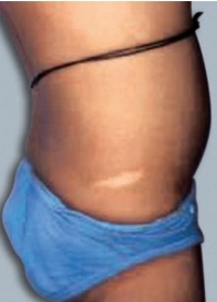
🔍 Key clinical features:
- ✓ Hypopigmented macule – lighter than surrounding skin
- ✓ Often ash-leaf or polygonal shape – sometimes confetti lesions
- ✓ Major diagnostic criterion – ≥3 macules, ≥5 mm diameter
- ✓ Best seen with Wood lamp – enhances contrast
- ✓ Present from birth/early infancy – often first sign of TSC
📋 Clinical scenario (examiner prompt)
A 3‑year‑old child is brought for a routine well-child visit. The mother has noticed several light-colored spots on the child's trunk that have been present since birth. On examination, there are ≥5 well-defined, hypopigmented macules ranging from 1‑3 cm in size on the trunk and extremities, with an ash-leaf or polygonal shape. The child is otherwise healthy with a normal neurologic examination. A Wood lamp examination enhances the visibility of the lesions. The mother has no similar lesions.
🧑⚕️ Examiner tasks · TOACS
1. Identify the diagnosis from the clinical image and context.
2. Describe the clinical features (hypopigmented ash-leaf macules, Wood lamp enhancement).
3. Explain the underlying condition (tuberous sclerosis complex, TSC1/TSC2, mTOR).
4. Discuss diagnostic criteria and management (major/minor criteria, screening, surveillance).
🎯 Expected answers (for examiners)
- • Diagnosis: Tuberous sclerosis complex – hypopigmented macules (ash-leaf spots)
- • Clinical features: Hypopigmented macules (usually >5mm, ash-leaf or polygonal shape), best seen under Wood lamp; present from birth or early infancy; may be confused with vitiligo, nevus depigmentosus
- • Genetics: TSC1 (9q34, hamartin) or TSC2 (16p13, tuberin) → mTOR overactivation; autosomal dominant, 2/3 de novo
- • Diagnostic criteria (major): ≥3 hypopigmented macules ≥5mm; facial angiofibromas; shagreen patch; ungual fibromas; cortical tubers; subependymal nodules; SEGA; cardiac rhabdomyoma; renal angiomyolipoma; LAM
- • Surveillance: MRI brain every 1-3y (SEGA); renal ultrasound every 1-3y; ophthalmology; echocardiogram; EEG; developmental screening
- • Treatment: Vigabatrin for infantile spasms; everolimus for SEGA, renal AML, refractory seizures
• Location: Trunk, extremities (anywhere)
• Appearance: Well-defined, hypopigmented, ash-leaf or polygonal shape
• Size: ≥3 macules, ≥5 mm diameter = major criterion
• Detection: Wood lamp enhances visibility
• Onset: Present at birth or early infancy
• Differential: Nevus depigmentosus, vitiligo (acquired), tuberous sclerosis
• Management: Diagnosis of TSC requires full evaluation and surveillance
⚡ Quick FCPS‑style MCQ
A 6-month-old infant has ≥5 hypopigmented ash-leaf macules on the trunk, best seen under Wood lamp. The most likely diagnosis is:
A. Vitiligo B. Tuberous sclerosis complex C. Nevus depigmentosus D. Pityriasis alba📌 Topic summary · Hypopigmented Macules in Tuberous Sclerosis
Hypopigmented macules >5mm
≥3 ash-leaf spots
Wood lamp enhances contrast
TSC1 (hamartin) or TSC2 (tuberin)
Seizures, cardiac rhabdomyoma, SEGA
Surveillance, everolimus, vigabatrin
| Feature | Tuberous Sclerosis – Hypopigmented Macules |
|---|---|
| Definition | Hypopigmented macules (ash-leaf spots) – areas of decreased melanin |
| Size criterion | ≥3 macules, ≥5 mm in diameter (major diagnostic feature) |
| Shape | Ash-leaf (polygonal) or confetti pattern |
| Detection | Best visualized with Wood lamp (ultraviolet) – enhances contrast |
| Onset | Present at birth or early infancy (often first sign) |
| Genetics | TSC1 (hamartin, 9q34) or TSC2 (tuberin, 16p13) → mTOR overactivation |
| Inheritance | Autosomal dominant, 2/3 de novo |
| Associated features | Seizures (infantile spasms), cardiac rhabdomyoma, renal angiomyolipoma, SEGA, facial angiofibromas, shagreen patch |
| Management | Diagnosis → MRI brain, renal US, echocardiogram, ophthalmology, EEG; vigabatrin for spasms; everolimus for SEGA/AML |