🧬 TOACS FCPS Station · Plexiform Neurofibroma in Neurofibromatosis
📷 Clinical Photograph – Plexiform Neurofibroma
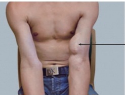
🔍 Key clinical features:
- ✓ Diffuse, infiltrating mass – often present at birth or early childhood
- ✓ Soft, "bag of worms" texture – palpable along nerve bundles
- ✓ May involve any nerve – commonly cervical, facial, lumbosacral, mediastinal
- ✓ Hyperpigmentation (cafe-au-lait) – overlying skin
- ✓ Can cause disfigurement, pain, functional impairment
- ✓ Risk of malignant transformation – to MPNST (malignant peripheral nerve sheath tumor)
📋 Clinical scenario (examiner prompt)
A 9‑year‑old child with a known diagnosis of neurofibromatosis type 1 (NF1) is brought in due to a rapidly enlarging mass on the left arm The mother reports that the mass has been present since birth but has increased in size over the past 3 months and is now associated with severe pain and limited movement. On examination, there is a large, diffuse, soft, infiltrating mass extending from the arm to the forearm, with overlying hyperpigmented skin. The child has multiple cafe‑au‑lait spots and axillary freckling.
🧑⚕️ Examiner tasks · TOACS
1. Identify the diagnosis from the clinical image and context.
2. Describe the clinical features (plexiform neurofibroma – diffuse, infiltrating mass, soft consistency, overlying hyperpigmentation).
3. Explain the underlying condition (NF1, NF1 gene mutation, neurofibromin, Ras pathway).
4. Discuss complications and management (MPNST risk, imaging, selumetinib, surgery, surveillance).
🎯 Expected answers (for examiners)
- • Diagnosis: Plexiform neurofibroma in NF1 (with concern for malignant transformation to MPNST)
- • Clinical features: Diffuse, infiltrating, soft "bag of worms" mass along nerve; often present at birth; overlying skin hyperpigmentation (CAL); may cause pain, disfigurement, functional impairment
- • NF1 diagnosis: ≥2 of: CALs (≥6), axillary/inguinal freckling, neurofibromas (≥2) or 1 plexiform, optic glioma, Lisch nodules, sphenoid dysplasia/tibial pseudarthrosis, first-degree relative, NF1 pathogenic variant
- • MPNST suspicion: Rapid growth, new or worsening pain, neurologic deficit, PET/CT uptake (SUVmax >3)
- • Imaging: MRI (to assess extent, infiltration), PET/CT (for malignant transformation)
- • Treatment: Selumetinib (MEK inhibitor) for inoperable symptomatic plexiform neurofibromas; surgical resection if feasible; chemotherapy for MPNST
- • Surveillance: Annual ophthalmology, BP monitoring, developmental screening, and routine physical exam for new/changing lesions
• Incidence: ~30% of NF1 patients, usually congenital
• Location: Head/neck (20%), trunk, extremities, lumbosacral
• Risk of MPNST: 8-13% lifetime risk (most common cause of death in NF1)
• Red flags: Rapid growth, persistent pain, neurologic deficit
• FDA-approved therapy: Selumetinib (MEK inhibitor) for inoperable symptomatic lesions
• Surgical considerations: Complete resection often difficult (infiltrating); debulking for cosmesis or symptom relief
⚡ Quick FCPS‑style MCQ
A 6-year-old with NF1 presents with a rapidly enlarging, painful neck mass with overlying hyperpigmentation. The most concerning complication is:
A. Benign growth of cutaneous neurofibroma B. Malignant transformation to MPNST C. Spontaneous regression D. Reactive lymphadenopathy📌 Topic summary · Plexiform Neurofibroma in NF1
Diffuse, infiltrating nerve sheath tumor
Neurofibromatosis type 1 (NF1)
Malignant transformation to MPNST (8-13%)
Rapid growth, pain, neurologic deficit
MRI, PET/CT for malignancy
Selumetinib, surgery, surveillance
| Feature | Plexiform Neurofibroma |
|---|---|
| Onset | Congenital (present at birth or early childhood) |
| Appearance | Diffuse, infiltrating, soft ("bag of worms"), overlying CAL |
| NF1 association | Diagnostic criterion (≥1 plexiform neurofibroma) |
| MPNST risk | 8-13% lifetime risk; most common cause of death in NF1 |
| Imaging | MRI for extent; PET/CT (SUVmax >3 suggestive of malignancy) |
| Treatment | Selumetinib (MEK inhibitor) for inoperable symptomatic lesions; surgical resection (partial/debulking); MPNST: chemotherapy, radiation, surgery |
| Surveillance | Annual ophthalmology, BP, developmental screening, physical exam; MRI if symptoms |