🧬 TOACS FCPS Station · Telangiectasia of Eyes in Ataxia-Telangiectasia
📷 Clinical Photograph – Ocular Telangiectasias
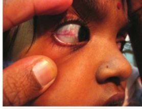
🔍 Key clinical features:
- ✓ Dilated, tortuous blood vessels on bulbar conjunctivae
- ✓ Oculocutaneous telangiectasias – also on sun-exposed skin (ears, nose, neck)
- ✓ Appear between ages 3-6 years – often the diagnostic clue
- ✓ Associated with progressive cerebellar ataxia – usually precedes telangiectasias
- ✓ Pathognomonic for ataxia-telangiectasia (Louis-Bar syndrome)
📋 Clinical scenario (examiner prompt)
A 5‑year‑old child is referred to the neurology clinic for gait instability and frequent falls. The parents report that the child has been unsteady since age 3 and has slurred speech. On examination, the child has truncal ataxia, dysarthria, and dilated, tortuous blood vessels on the bulbar conjunctivae of both eyes. The child has a history of recurrent sinopulmonary infections and low IgA on a previous workup.
🧑⚕️ Examiner tasks · TOACS
1. Identify the diagnosis from the clinical image and context.
2. Describe the clinical features (ocular telangiectasias, cerebellar ataxia, immunodeficiency).
3. Explain the underlying condition (ataxia-telangiectasia, ATM gene, DNA repair defect).
4. Discuss diagnosis and management (AFP, immunoglobulins, avoid radiation, supportive care).
🎯 Expected answers (for examiners)
- • Diagnosis: Ataxia-telangiectasia (Louis-Bar syndrome)
- • Clinical features: Oculocutaneous telangiectasias (bulbar conjunctivae, sun-exposed skin), progressive cerebellar ataxia (onset 1-4 years, wheelchair by adolescence), dysarthria, oculomotor apraxia, choreoathetosis, dystonia; immunodeficiency (selective IgA deficiency, low IgG2, T-cell lymphopenia, poor antibody responses); recurrent sinopulmonary infections
- • Genetics: ATM gene (11q22) – autosomal recessive; defective DNA double-strand break repair and cell cycle checkpoint
- • Diagnostic tests: Elevated serum alpha-fetoprotein (AFP) in >95% after 2 years; low IgA, low IgG2, lymphopenia; ATM sequencing; chromosomal breakage (radiosensitivity)
- • Management: Supportive care (PT/OT, speech therapy, feeding tube); IVIG for severe infections; avoid live vaccines; avoid ionizing radiation (extreme radiosensitivity); monitor for malignancy (lymphoma, leukemia); genetic counseling
• Location: Bulbar conjunctivae (dilated, tortuous vessels)
• Onset: Ages 3-6 years (later than ataxia)
• Pathognomonic for ataxia-telangiectasia
• Associated: Ataxia, dysarthria, immunodeficiency, elevated AFP
• Management: Supportive, avoid radiation, treat infections
• Prognosis: Progressive; death from pulmonary failure or malignancy
⚡ Quick FCPS‑style MCQ
A 5-year-old with progressive ataxia, dysarthria, and dilated blood vessels on the bulbar conjunctivae. The most characteristic laboratory finding is:
A. Elevated alpha-fetoprotein (AFP) B. Low C3 C. Absent B cells D. Positive antinuclear antibody📌 Topic summary · Ocular Telangiectasias in Ataxia-Telangiectasia
Ataxia-telangiectasia (Louis-Bar)
Dilated conjunctival vessels
ATM (11q22)
Elevated AFP, IgA deficiency
Extreme – avoid radiation
Supportive, IVIG, PT/OT
| Feature | Ataxia-Telangiectasia |
|---|---|
| Gene | ATM (ataxia-telangiectasia mutated) – 11q22 |
| Inheritance | Autosomal recessive |
| Ocular telangiectasias | Dilated, tortuous vessels on bulbar conjunctivae; appear ages 3-6 years; pathognomonic |
| Neurologic | Progressive cerebellar ataxia (onset 1-4 years), dysarthria, oculomotor apraxia, dystonia, choreoathetosis |
| Immunodeficiency | Selective IgA deficiency (50-80%), low IgG2, progressive T-cell lymphopenia, poor antibody responses |
| Laboratory | Elevated AFP (>95% after 2 years), low IgA, lymphopenia, elevated chromosomal breakage |
| Radiosensitivity | Extreme – avoid X-rays, CT, radiotherapy; use MRI |
| Malignancy risk | Lymphoma, leukemia, breast cancer (carriers); 10-30% lifetime risk |
| Management | Supportive (PT/OT, speech, feeding tube), IVIG for infections, avoid live vaccines, treat infections aggressively, cancer surveillance |
| Prognosis | Progressive; median survival 20-30 years; death from pulmonary failure or malignancy |