🍄 TOACS FCPS Station · Annular Rash in Tinea faciei
📷 Clinical Photograph – Annular Rash (Tinea faciei)
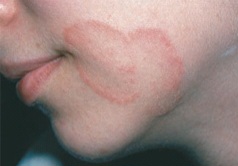
🔍 Key clinical features:
- ✓ Annular (ring-shaped) plaque – well-demarcated
- ✓ Central clearing – classic 'ringworm' appearance
- ✓ Active scaly border – erythematous, elevated edge
- ✓ Mild pruritus – variable
- ✓ Can affect any body surface – trunk, extremities, face
📋 Clinical scenario (examiner prompt)
A 7‑year‑old child is brought in with a ring-shaped rash on the face that has been slowly enlarging over the past few weeks. The lesion is erythematous with a scaly, elevated border and central clearing. The child reports mild itching. The family has a new kitten at home. The child is otherwise healthy and has no other skin lesions.
🧑⚕️ Examiner tasks · TOACS
1. Identify the diagnosis from the clinical image and context.
2. Describe the clinical features (annular plaque, central clearing, active scaly border).
3. Explain the etiology and transmission (dermatophytes, contact with animals/fomites).
4. Discuss diagnosis and management (KOH prep, topical antifungals, when to use systemic).
🎯 Expected answers (for examiners)
- • Diagnosis: Tinea Corporis (dermatophyte infection, ringworm)
- • Clinical features: Annular (ring-shaped) erythematous plaque with central clearing, active scaly border (elevated, erythematous), mild pruritus, slowly enlarging
- • Etiology: Dermatophytes: Trichophyton rubrum (most common), Microsporum canis (from animals), Epidermophyton floccosum; transmission via direct contact with infected humans, animals, or fomites
- • Diagnosis: Clinical; KOH prep (septate hyphae, branching); Wood lamp (green fluorescence for Microsporum canis); fungal culture (definitive)
- • Treatment: Topical antifungals: clotrimazole, miconazole, ketoconazole, terbinafine (BID) for 2-4 weeks. Systemic therapy: terbinafine or griseofulvin for extensive disease, immunosuppressed, or Majocchi granuloma
- • Differential: Granuloma annulare (no scale), nummular eczema, pityriasis rosea, psoriasis, Lyme disease (erythema migrans)
• Appearance: Annular plaque with central clearing and active scaly border
• Pathogens: T. rubrum (human), M. canis (animal), E. floccosum
• Diagnosis: KOH prep (septate hyphae), culture
• Treatment: Topical antifungals (2-4 weeks)
• Systemic indication: Extensive, resistant, Majocchi granuloma
• Differential: Granuloma annulare (no scale), nummular eczema
⚡ Quick FCPS‑style MCQ
A 7-year-old with a ring-shaped, erythematous plaque with central clearing and an active scaly border on the face. The most likely diagnosis and first-line treatment are:
A. Granuloma annulare – topical steroids B. Tinea faciei – topical antifungal (clotrimazole/terbinafine) C. Nummular eczema – emollients D. Psoriasis – topical corticosteroids📌 Topic summary · Annular Rash in Tinea faciei
Dermatophyte infection of glabrous skin
Annular plaque with central clearing
T. rubrum, M. canis, E. floccosum
KOH prep (septate hyphae)
Topical antifungals (2-4 weeks)
Extensive, resistant, Majocchi granuloma
| Feature | Tinea faciei |
|---|---|
| Definition | Superficial fungal infection (dermatophytosis) of the glabrous (non-hair-bearing) skin |
| Clinical appearance | Annular (ring-shaped) erythematous plaque with central clearing and active scaly border; may be single or multiple |
| Common pathogens | Trichophyton rubrum, Microsporum canis (animals), Epidermophyton floccosum |
| Transmission | Direct contact with infected humans, animals (kittens, puppies), or fomites (towels, clothing, sports equipment) |
| Diagnosis | KOH prep (septate hyphae, branching); Wood lamp (green fluorescence for M. canis); fungal culture |
| Treatment – Topical | Azoles (clotrimazole, miconazole, ketoconazole) or terbinafine cream BID for 2-4 weeks |
| Treatment – Systemic | Terbinafine (250 mg daily) or griseofulvin for extensive, resistant, or deep infection (Majocchi granuloma) |
| Differential diagnosis | Granuloma annulare (no scale), nummular eczema, pityriasis rosea, psoriasis, erythema migrans (Lyme disease) |