🩻 TOACS FCPS Station · Chest X‑ray: Right Superior Basal Segment Consolidation
📷 Chest X‑ray (PA view)
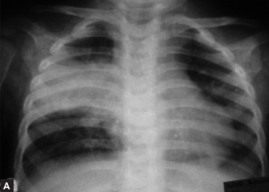
🔍 Key radiographic features:
- ✓ Homogeneous opacification (consolidation) in right superior basal segment
- ✓ Air bronchogram – air-filled bronchi visible within the opacified lung
- ✓ Lobar distribution – typical of bacterial pneumonia (S. pneumoniae)
- ✓ No loss of lung volume (unlike atelectasis)
- ✓ Sharply defined borders at fissures (if lobar)
📋 Clinical scenario (examiner prompt)
A 4‑year‑old child presents with fever (39.5°C), productive cough, and tachypnoea (respiratory rate 44/min). He has decreased breath sounds and crackles in the right lower chest. Oxygen saturation is 94% on room air. The chest X‑ray (PA view) shows consolidation in the right superior basal segment with air bronchograms.
🧑⚕️ Examiner tasks · TOACS
1. Identify the diagnosis from the X‑ray and clinical context.
2. Describe the pathology (lobar consolidation/airspace disease).
3. Describe the X‑ray findings (consolidation, air bronchogram, segmental distribution).
4. Discuss management (outpatient vs inpatient, antibiotics).
🎯 Expected answers (for examiners)
- • Diagnosis: Community-Acquired Pneumonia (CAP) – bacterial (lobar) pneumonia
- • Pathology: Alveolar filling (consolidation) with inflammatory exudate; most common: S. pneumoniae
- • X‑ray: Homogeneous opacification, air bronchogram, right superior basal segment
- • Management: Outpatient amoxicillin 90 mg/kg/day if mild; admit if hypoxemic or respiratory distress
⚡ Quick FCPS‑style MCQ
A 4‑year‑old with lobar consolidation and air bronchogram. First‑line outpatient antibiotic is:
A. Amoxicillin 90 mg/kg/day B. Azithromycin C. Ceftriaxone D. Vancomycin📌 Topic summary ·
Community‑Acquired Pneumonia (CAP)
S. pneumoniae, H. influenzae, S. aureus
Consolidation, air bronchogram, lobar
Amoxicillin 90 mg/kg/day
Ampicillin or ceftriaxone
M. pneumoniae – macrolide
| Age | Common Pathogens | Empiric Antibiotic |
|---|---|---|
| Neonate (<3 weeks) | GBS, E. coli, Listeria | Ampicillin + gentamicin/cefotaxime |
| 3 weeks - 3 months | RSV, Chlamydia, S. pneumoniae | Macrolide (if afebrile) or ampicillin/ceftriaxone |
| 4 months - 4 years | RSV, S. pneumoniae, H. influenzae | Amoxicillin (outpatient) / ampicillin or ceftriaxone (inpatient) |
| ≥5 years | M. pneumoniae, S. pneumoniae, C. pneumoniae | Amoxicillin or macrolide |