🩻 TOACS FCPS Station · X‑ray Hip: Epiphyseal Dysgenesis (Congenital Hypothyroidism)
📷 X‑ray Hip (AP view)
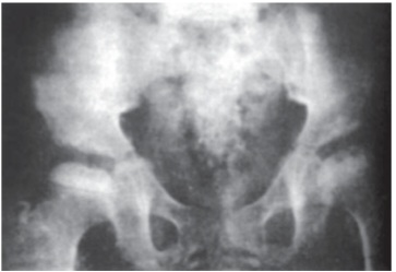
🔍 Key radiographic features:
- ✓ Delayed appearance of the femoral head epiphysis (absent or small)
- ✓ Fragmented, stippled appearance – multiple small ossification centers
- ✓ Irregular epiphyseal contour – “moth-eaten” appearance
- ✓ Widened joint space – due to delayed ossification
- ✓ Delayed bone age – overall skeletal maturation retarded
📋 Clinical scenario (examiner prompt)
A 9‑month‑old infant with known congenital hypothyroidism (diagnosed by newborn screening) presents for routine follow‑up. The infant has been on levothyroxine since day 14 of life but has had poor adherence to therapy. On examination, there is hypotonia, macroglossia, and delayed motor milestones. A hip X‑ray (AP view) is shown.
🧑⚕️ Examiner tasks · TOACS
1. Identify the diagnosis from the hip X‑ray and clinical context.
2. Describe the X‑ray findings (epiphyseal dysgenesis, fragmented femoral head).
3. Explain the pathophysiology (thyroid hormone deficiency → impaired ossification).
4. Discuss management (levothyroxine adherence, monitoring, prognosis).
🎯 Expected answers (for examiners)
- • Diagnosis: Epiphyseal dysgenesis due to congenital hypothyroidism (poorly controlled)
- • X‑ray findings: Fragmented/stippled femoral head epiphysis, delayed ossification, widened joint space
- • Pathophysiology: Thyroid hormone deficiency → impaired chondrocyte maturation and endochondral ossification
- • Management: Optimize levothyroxine dose (10-15 mcg/kg/day), ensure adherence, monitor TSH/free T4, repeat bone age
⚡ Quick FCPS‑style MCQ
A fragmented, stippled femoral head epiphysis on hip X‑ray in an infant with congenital hypothyroidism is due to:
A. Thyroid hormone deficiency → impaired endochondral ossification B. Growth hormone deficiency C. Vitamin D deficiency D. Osteomyelitis📌 Topic summary ·
Fragmented, stippled epiphysis
Femoral head (hip)
Impaired endochondral ossification
Delayed bone age
Levothyroxine (10-15 mcg/kg/day)
Reversible with treatment
| Feature | Congenital Hypothyroidism |
|---|---|
| Bone age | Delayed (retarded skeletal maturation) |
| Epiphyses | Fragmented, stippled, absent (dysgenesis) |
| Skull | Wide sutures, large fontanels |
| Reversibility | Reversible with adequate levothyroxine |