🩻 TOACS FCPS Station · X‑ray Long Bones: Osteogenesis Imperfecta (OI)
📷 X‑ray Long Bones (AP view)
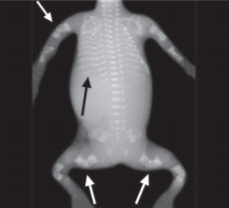
🔍 Key radiographic features:
- ✓ Multiple fractures – healing and healed fractures at various stages
- ✓ Bowed long bones – femoral and tibial bowing (especially in Type III)
- ✓ Osteopenia – decreased bone density with thin cortices
- ✓ Popcorn calcifications – metaphyseal areas (Type III)
- ✓ Wormian bones – intrasutural bones (skull X‑ray)
📋 Clinical scenario (examiner prompt)
A 1‑month‑old child presents with a history of multiple fractures from minimal trauma, bowed legs, and short stature. On examination, there are blue sclerae and dentinogenesis imperfecta. A long bone X‑ray (AP view) is shown.
🧑⚕️ Examiner tasks · TOACS
1. Identify the diagnosis from the X‑ray and clinical context.
2. Describe the X‑ray findings (fractures, bowed bones, osteopenia).
3. Explain the pathophysiology (COL1A1/COL1A2 mutation → defective collagen).
4. Discuss classification and management (Sillence types, bisphosphonates, rodding).
🎯 Expected answers (for examiners)
- • Diagnosis: Osteogenesis imperfecta (likely Type III or IV)
- • X‑ray findings: Multiple fractures (healing/healed), bowed long bones, osteopenia
- • Pathophysiology: Defective type I collagen (COL1A1/COL1A2) → bone fragility
- • Management: IV pamidronate (bisphosphonates), physical therapy, intramedullary rodding, genetic counseling
⚡ Quick FCPS‑style MCQ
A child with bowed femurs, multiple fractures, blue sclerae, and dentinogenesis imperfecta most likely has:
A. Osteogenesis imperfecta Type I B. Osteogenesis imperfecta Type III C. Rickets D. Hypophosphatasia📌 Topic summary ·
Blue sclerae, mild fractures
Perinatal lethal, crumpled femurs
White sclerae, progressive deformity
White sclerae, moderate severity
IV pamidronate (bisphosphonate)
Intramedullary telescoping rods
| OI Type | Sclerae | Severity | Key Feature |
|---|---|---|---|
| Type I | Blue | Mild | Hearing loss, fractures decrease after puberty |
| Type II | Blue | Lethal | Crumpled femurs, beaded ribs |
| Type III | White | Severe | Bowed femurs, triangular facies, popcorn calcifications |
| Type IV | White | Moderate | Variable short stature |